Signs of degenerative changes in the menisci. Degenerative changes in the knee meniscus and types of pathologies. Features of damage to the internal meniscus
The human body is often compared to cars: the heart is the engine, the stomach is the fuel tank, and the brain sets the entire device in motion. Where are the shock absorbers in humans? Of course, in places that are experiencing increased stress: between the vertebrae there are cartilaginous discs, and in the knee joint there are two “shock absorbers” - menisci. Lateral (external) and medial (internal). Although the results of degenerative changes in the menisci will not stop the activity of the body as a whole, they will deliver a lot of unpleasant sensations for sure.

What are meniscus degenerative changes?
Degenerative changes are anatomical damage to an organ resulting from trauma, atypical joint structure or disease. Meniscus degeneration is most often the result of an injury, sometimes not even obvious: one unsuccessful turn of the lower leg can cause damage to the cartilaginous disc, which is accompanied by severe pain.
Most often, due to the anatomical structure, the medial meniscus undergoes degeneration. If the external cartilage, which cushions the movement of the knee joint, does not have rigid fixation and is displaced to either side if necessary, then the medial cartilage is rigidly fixed in the joint, and its horns are in the immediate vicinity of the condyles. One sharp turn of the lower leg - and the meniscus does not have time to escape from the displaced process of the bone, the result is damage or rupture.
Degenerative changes can be different:
- separation from the attachment point;
- rupture of the horns and the body of the meniscus;
- excessive mobility as a result of rupture of intermenis ligaments;
- cyst - the formation of cavities inside the cartilage filled with fluid;
- Meniscopathies - dystrophic changes that develop under the influence of minor injuries, as well as a complication of gout, osteoarthritis, rheumatism, tuberculosis and a number of other diseases.

Typical symptoms
If you are haunted by aching knee pain, which sometimes disappears, then appears with renewed vigor, you can already assume the presence of changes in the meniscus. About 90% of the pathologies of the knee joint are caused by damage to the "shock absorber".
Symptoms largely depend on the nature of the pathology. The tears are accompanied by severe soreness, blockage of the leg in a bent state, and swelling. With serious damage to the medial meniscus, hemorrhage often occurs in the articular cavity - hemarthrosis. Cystosis of the meniscus is also characterized by significant swelling and severe pain.
Tears, detachments from the attachment site are often chronic in nature and are manifested by the periodic appearance of pain and a feeling of hindrance in movement.
There is such a diagnostic test: go up and down a ladder or slope. With pathology of the meniscus, when moving downward, the pain in the knee increases.
Chronic course is also characterized by secondary degenerative-dystrophic transformations in the medial meniscus, that is, arising from other pathologies of the body or diseases. Often in such cases, clicks and rolling * of the joint in motion after prolonged rest are noted, sometimes pain in the knees appears. The increase in symptoms occurs gradually as the cartilage layer becomes thinner and the accumulation of salts or crystals of uric acid in it (the latter - with gout). In the absence of adequate treatment, the final stage of meniscopathy is contracture - a stable violation (limitation) of joint mobility.
* Rolling - sensations of pathological mobility, instability and displacement of the articular surfaces of the bones.
The following symptoms are common to all types of meniscus degeneration:
- soreness
- swelling
- blockage of a joint in a bent position or sensation of a foreign body in the knee,
- clicks and crunch,
- swelling of the knees with a long absence of movement.

Degeneration causes
The anatomical features of the location and structure of the menisci determine the high incidence of pathologies both among young people and among people of mature age. Most often, athletes, ballerinas, dancers - that is, people who are in constant motion and experiencing high loads - suffer from tears, injuries and cystosis.
Other possible reasons:
- dysplasia - abnormal formation of the knee joint;
- gout, syphilis, tuberculosis, rheumatism and other diseases that can affect the joints;
- sprains of the ligaments, as well as their incorrect formation;
- flat feet (low amortization of the foot is compensated by increased load on the knee);
- high physical activity;
- excess weight.
Diagnostics
In case of acute injuries of the menisci, there is usually no doubt - a knee block in a characteristic position, pain and clicks during straightening make it possible to establish the correct diagnosis in 90% of cases.
Degenerative-dystrophic transformations can not always be determined during examination due to the absence of vivid symptoms and, often, a positive reaction to special tests. In such cases, they resort to instrumental research methods:
- MRI allows you to obtain a volumetric image of all tissues of the knee: the articular surfaces of the bones, the ligamentous apparatus and the joint itself.
- During arthroscopy, an endoscope is inserted through a miniature incision into the articular cavity, with the help of which the condition of the tissues and synovial fluid is monitored (on the monitor).
Treatment methods
Therapy for degenerative changes in the menisci depends entirely on the nature of the damage. Acute injuries are a direct indication for the use of conservative treatment methods:
- First of all, a puncture of the joint is performed, eliminating its swelling and restoring mobility. Sometimes several procedures are required, since active exudation (release of inflammatory fluid) in the joint lasts up to three to four days.
- Analgesics are prescribed, preference is given to narcotic drugs (Promedol and its derivatives), because other drugs in this case, as a rule, are not able to relieve the patient of pain.
- Chondroprotectors provide the body with the necessary substances to restore the damaged area of the meniscus.
- Anti-inflammatory drugs.
- At the stage of rehabilitation, physiotherapeutic methods are used as an auxiliary means - ozokerite, UHF, iontophoresis, shock wave therapy.
- For 14 days, a splint is applied to the straightened leg, which ensures the fixation of the joint in the required position.
In case of ruptures, surgical intervention is indicated: through two miniature incisions, instruments are inserted into the knee joint and the damaged area is sutured. Serious injury may necessitate the removal of the cartilage lining of the joint and replacement with an artificial one. All surgical manipulations are performed only after the signs of inflammation have faded away.
Chronic dystrophies, joint dysplasia and abnormal development of the ligamentous apparatus require only surgical treatment.
If the cause of degeneration is chronic diseases such as rheumatism and gout - along with surgical methods, treatment of the underlying disease is also performed (diets, immunocorrectors and other methods).
Degenerative transformations of the menisci is a fairly common pathology that requires an immediate visit to a specialist. The functioning of the joint in the future depends on the timeliness of treatment, and delays can cause the spread of degenerative processes to the rest of the elements of the joint. Therefore, do not postpone your visit to the doctor, take care of yourself and be healthy!
Degenerative changes in the meniscus are its anatomical damage that occurs after injury, a previous illness or an atypical structure of the joints. Most often, pathological changes in the menisci occur as a result of trauma, when the cartilaginous disc is damaged and provokes attacks of pain. Degenerative damage to the internal meniscus occurs more often in men than in women. It manifests itself in almost half of the cases.
Table of contents [Show]
General information
The human body is an extremely complex mechanism and its work must always be adjusted. Articular cartilage acts as shock absorbers that normalize and facilitate joint mobility. Cartilage tissue, located in the knee joint in the form of menisci, helps to reduce surface friction, improve joint rotation and limit mobility. There are two menisci in the knee joint: external (lateral) and internal (medial).
Degenerative changes in the cartilage lining of the knee joint are characteristic lesions that are the result of injuries (often in athletes), they can be complicated by the course of the disease or structural features of the joint. Among all joint diseases, degenerative changes in the menisci are in the first place.
Signs of change are:
- rupture of the horns and the body of the meniscus itself;
- the formation of a hollow cyst that is filled with fluid;
- the development of meniscopathy, the process of degeneration, which occurs as a result of rheumatism, tuberculosis;
- detachment of cartilage;
- rupture of the ligaments that connect the menisci.
The meniscus is the cartilaginous layer inside the knee joint, which mainly performs a shock-absorbing function. Tears of joint spacers can occur after injuries that occur in young people during physical exertion, and can also be degenerative, which occur in the elderly and can develop without injury against the background of degenerative changes in the meniscus, which are a variant of the course of arthrosis of the knee.
Lack of treatment for a traumatic rupture can lead to the fact that it subsequently becomes a chronic pathology.
To diagnose a meniscus rupture, it is necessary to conduct an ultrasound scan, MRI. Meniscus tears can be found in the anterior horn, posterior horn, and in the body of the meniscus. Damage to the meniscus can lead to mechanical obstruction of movement, and cause pain syndromes.
The dangling part of the meniscus provokes the destruction of the adjacent cartilage.
With a traumatic rupture of the meniscus, swelling of the knee joint and pain occurs. If the rupture occurs in a place where there are blood vessels, then hemarthrosis occurs. It is manifested by swelling above the kneecap. If the cartilage lining is damaged, the part that comes off and dangles can interfere with free movement in the knee. Small tears can cause painful clicks or a feeling of stiffness. In case of large ruptures, blockage of the joint may occur due to the fact that the torn fragment of the cartilaginous lining is moved to the center of the joint and provokes "jamming" of the joint.
When the posterior horn of the meniscus is torn, the flexion process is limited; when the body of the meniscus or its anterior horn is torn, pain occurs during the extension process in the knee joint. Pain syndromes with a rupture of the posterior horn of the meniscus can be so severe that it is impossible to step on the leg, and sometimes the rupture of the meniscus is manifested only by pain when making certain movements.
With an acute tear of the anterior cruciate ligament, edema may develop faster and be more pronounced. Damage to the lateral cartilage pad also occurs. Degenerative cartilage tears can occur with the slightest physical exertion, especially when it comes to the older generation. With a degenerative rupture of the medial meniscus, the adjacent cartilage, which covers more of the tibia and femur, is often damaged.
General symptoms of cartilage damage:
- clicks and crunching;
- swelling;
- soreness;
- with a long stay in one position, knees flow;
- joint blockage with bent knees.
Causes of meniscus lesions
The structure and anatomical features of the location of the menisci cause a high incidence of pathologies in different age categories. At risk are athletes who are prone to tears, injuries and cysts.
Possible causes of ruptured cartilage lining:
- improper formation or sprain of the ligaments;
- flat feet;
- malformed knee joint;
- the presence of gout, syphilis, tuberculosis, rheumatism and other diseases that can affect the joints;
- excess weight.
Forms of the disease
Damage to the external meniscus of the knee joint.
Trauma to the lateral meniscus in adults is extremely rare. This happens more often with children and adolescents. As a consequence of such an injury, blockade is rare.
Symptoms of damage to the lateral meniscus include:
- pain syndromes in the tissues in the area of the collateral ligament;
- pronounced synovitis;
- unpleasant sensation of pain in the area of the peroneal ligament;
- low tone in the muscles of the front of the thigh.
In the event of a rupture of the external cartilage, the knee joint can be bent at a right angle and the patient himself can unblock it. In general, the signs of this injury are not very pronounced. Diagnosing such an injury is quite problematic due to the inconsistent pain sensations. A congenital developmental anomaly is possible - a continuous (disc-shaped) external meniscus. It can be easily confused with cartilage tear. With this pathology, the cartilage has the shape of a disc. Signs of a solid external meniscus can appear during adolescence and can also be found at an older age.
Damage to the medial meniscus of the knee
A common injury to the medial meniscus is rupture. Basically, the rupture of the middle part occurs, while the ends remain intact.
There are three types of damage to the medial meniscus:
- rupture of the ligament that fixes the internal organ;
- rupture of the cartilage itself;
- rupture of cartilage tissue.
Detachment with pinching of the anterior horn of the inner meniscus provokes a blockage of the knee joint, which does not cause flexion of the knee. This phenomenon is temporary, since unblocking will restore movement in the joint. Damage to the posterior horn of the medial meniscus is a more serious injury. at the same time, blocking, jumping out and bending of the knee occurs.
Chronic degeneration and trauma of the menisci
The process of affecting the left and right cartilage to the same extent.
The causes of meniscus degeneration include:
- sharp extension of the leg;
- deposition of mucin in tissues;
- severe trauma;
- rheumatism;
- gout.
Diagnosis of the disease
Diagnosis of the disease can be carried out using the following studies:
- Magnetic resonance imaging;
- CT scan;
- X-ray;
- Diagnostic arthroscopy.
In order to reveal accurate diagnosis- meniscus rupture, you should consult a specialist. He needs to tell him under what circumstances you have pain. Any changes in the menisci are painful. On examination, the hip and knee joint are examined. With effusion, hemarthrosis or synovitis may be suspected.
Researching
X-ray is performed for any pain in the knee joint. It is carried out in the following projections:
- Side projection;
- Direct projection in a standing position, and when bending the knees at 45 °;
- Axial projection.
MRI - allows you to see the cartilage in several planes, to assess the condition of other periarticular and articular formations, which is important if there is doubt about the diagnosis. MRI in diagnosing meniscus problems has an accuracy of up to 95%. In the sagittal plane, the cartilaginous pad takes on the shape of a butterfly. When a rupture occurs, the symptom of a "double posterior cruciate ligament" occurs, when the meniscus is adjacent to the posterior cruciate ligament and is in the intercondylar fossa of the femur.
Treatment
After diagnostics and confirmation of the diagnosis, the specialist prescribes complex therapeutic methods, including a set of such measures:
- puncture from the knee joint;
- physiotherapy appointment: phonophoresis, UHF, iontophoresis, ozokerite;
- the appointment of analgesics, drugs containing narcotic substances (Promedol), NSAIDs, chondroprotectors (provide the body with substances that help restore the damaged area of the meniscus).
For 2 weeks, a splint is applied to the straightened leg, which provides fixation of the joint in the right position... In case of ruptures, chronic dystrophy, dysplasia of the joints, surgical intervention is performed. In the presence of gout or rheumatism, treatment of the underlying disease that provoked the process of degenerative changes is also carried out.
The main method of treating pathologies of knee cartilage is surgery. Arthroscopy is performed, the operation is performed through two incisions one centimeter long. The torn off part of the meniscus is removed, and its inner edge is leveled. After such an operation, the recovery period depends on the patient's condition, but on average it ranges from 2 days to several weeks.
A pair of cartilage pads present in the human knee joint are called menisci. Like the cartilaginous discs of the spine, menisci perform both shock-absorbing and stabilizing functions, protecting the joint from excessive movement and overuse.
To increase functionality in the human knee, there are two whole types of elastic formations:
- External (lateral).
- Internal (medial).
And although pathological changes in the meniscus do not pose a fatal threat, the patient's quality of life for any pathology, including a meniscus rupture, invariably falls.
Degenerative changes in the menisci - what is the danger
Degenerative changes are acquired anomalies of the anatomy of an organ caused by mechanical damage or any, possibly even unconscious, joint injury. Unlike the outer, more mobile and mobile meniscus, the inner one is rigidly connected to the lateral knee ligament, which often leads to injuries accompanied by severe pain.
The most common degenerative changes in cartilage are:
- Violation of the integrity of the ligament at the attachment points of both horns or the body in the paracapsular area.
- Transchondral tears of the body and horns of the meniscus.
- Meniscopathies of various etiologies, which lead to complications after infectious and a number of autoimmune diseases.
- Rupture of intermeniscus ligaments, leading to pronounced degenerative changes in the joint.
Clinical picture
The manifestations of the disease are quite diverse and directly depend on the nature of the damage. The following signs of degenerative changes can be considered as common:
- Pronounced pain syndrome.
- Distinctly audible crunching and single clicking sounds in the knee joint.
- Inability to fully extend the leg.
- The illusion of the presence of a foreign body in the knee.
- Puffiness
- Morning stiffness of movements, disappearing after attempts to disperse.
Causes of pathology
Most often, degenerative changes are observed among people who lead an active lifestyle and sufficiently load the knee joint.
In addition, anatomical damage can be caused by dysplasia, heterogeneous and infectious diseases of internal organs, injuries of the lower extremities and pathological changes in the shape of the foot.
Excess body weight and excessive physical activity can also provoke a violation of the integrity of the meniscus.
Diagnostics
In the absence of pronounced symptoms that clearly indicate the degenerative-dystrophic nature of the damage, the doctor prescribes a number of non-invasive research methods designed to clarify the diagnosis:
- MRI, thanks to which the doctor can see the affected area in different planes.
- Arthroscopy, which allows you to examine the damage from the inside using an endoscope inserted into the joint.
Therapy
Depending on the nature of the damage, the doctor chooses a variety of therapeutic measures, the task of which is to combat the manifestations of pathology.
If the signs of the disease are acute, conservative therapy is recommended. After the puncture, the doctor prescribes powerful pain relievers to reduce the symptoms of acute pain.
The next treatment stage includes the use of chondroprotectors, anti-inflammatory drugs and physiotherapy. Two weeks later, after the limb has partially regained its mobility, a splint is applied to it to fix the joint.
If the joint damage is in the nature of an injury, as well as in the case of a chronic manifestation of the disease, the best way treatment will be surgery.
In order to keep the knee joint healthy, it is recommended to get a qualified consultation from a Koleno21 specialist at the first signs of degenerative changes. This is the only way you can stop the attack and prevent its further spread.
Degenerative changes in the knee meniscus are common injuries in any age group. The injury is typical for athletes, but also ordinary people occurs quite often.
The onset of dystrophic changes leads to the disruption of the motor system. Accordingly, it is very important to undergo a course of treatment in a timely manner in order to prevent the consequences. That is why, when the first symptomatology occurs, you need to waste no time going to a doctor's consultation. The course of treatment takes a long time.
To fully understand the severity of the damage, you need to know what a meniscus is needed for. It is a cartilage tissue that acts as a shock absorber in the joint and also stabilizes the knee. The meniscus affects the improvement of the rotational movement of the entire joint.
Prerequisites of the disease
There are two menisci in the knee:
- internal, or lateral;
- external, or medial

Meniscus anatomy in the joint
Due to the peculiarities of the structure of the joint or genetic predisposition degenerative changes in the medial meniscus occur much more often, especially in the posterior horn. There are no arteries in the meniscus, so a comprehensive course of treatment is prescribed. With the onset of a disease on the knee, swelling immediately forms, pain begins to disturb.
The main causes of degeneration of the meniscus.
- Gradually happens joint wear, accordingly, it is simply impossible to avoid the problem of full knee work in the older age category.
- Changes in the knee are due to a sharp load on the joint when unbending. Moreover, a break is possible.
- The risk group includes people who perform every day physical exercise.
- The cause may be anatomical or genetic predisposition.
- Previously suffered inflammation of the knee joint. In cases of damage to the medial meniscus, the formation of a hollow cyst, filling with fluid, is observed.
Symptoms of the degenerative process
There are two types of damage:
The main symptomatology of changes in the posterior horn of the medial meniscus;

An acute form of degenerative injury to the knee meniscus can take a long time. Chronic appearance may occur 2 weeks after the rupture has occurred. Pain sensations become stronger and are permanent. The feeling of pain may not leave even during movement. Experts strongly recommend that you stay in bed so as not to injure your leg even more. When the patient begins to walk, a crunch or click may appear - this signals a joint disease. When probing a diseased part of the body, a sensation of an articular ridge is possible.
With degenerative damage to the medial meniscus in a chronic form, thinning of the cartilage tissue often occurs. General clinical manifestations when changing look like this:
- redness on the knee;
- strong painful sensations, which only intensify during walking;
- crunching sensation during movement.
Reasons for the development of changes
 Degenerative-dystrophic changes in the menisci can be diagnosed in a person at any age. The risk group includes people who go in for sports professionally. The gap occurs due to an incorrect sharp movement.
Among other reasons, the presence of flat feet, previously obtained sprains or incorrect formation of the knee joint, directly related to the destruction of joints, problems (syphilis, gout, tuberculosis, rheumatism, and others) are also distinguished. Main role plays the weight of a person's body. With excessive weight, the likelihood of pathology formation increases significantly.
Characteristics of traumatic and degenerative meniscus rupture
Degenerative rupture of the posterior horn of the medial meniscus is characterized by recurrent pain. This injury often occurs in older people. The patient has swelling of the injured knee. Given the severity of the change, the course of treatment lasts long enough. The patient is prescribed complex treatment, taking into account all individual characteristics, the degree of damage. It is strictly forbidden to self-medicate or resort to folk methods, it will not show the desired result, time will be wasted, and therefore complications may arise.
The rupture of the lateral meniscus of the knee joint is mainly traumatic in nature. The patient immediately begins to be bothered by severe pains, redness occurs, due to the fact that blood accumulates in the damaged area, and swelling of the knee occurs. Accordingly, the patient must immediately contact a doctor for help.
Diagnosis and treatment of a damaged knee joint
In cases where the patient has a degenerative-dystrophic rupture of the knee joint meniscus in an acute form, confirm the diagnosis quickly enough, due to the fact that at this time the symptomatology of the pathology has a pronounced character. Certain difficulties arise in the chronic form of pathology. In this situation, the patient is prescribed passing a specific examination.
The course of treatment is directly related to the severity of the injury. Degenerative changes in the medial meniscus are treated with conservative ways... In cases where the patient has an accumulation of blood in a part of the affected joint, he is prescribed strict bed rest, complete exclusion of loads on the injured knee. Typically, the course of treatment lasts from 1 to a month. The patient is prescribed special physiotherapy procedures, as well as physiotherapy exercises, all exercises are performed under the supervision of specialists.
It must be remembered that all procedures and exercises begin to be performed only when the inflammatory process is eliminated. In situations where conservative treatment did not show the desired result or the patient had a ruptured internal meniscus, it is prescribed surgical intervention, since the operation is the only way to help.
June 12, 2017 No comments
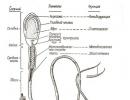
Menisci are crescent-shaped layers of cartilage within the knee joint between the surfaces of the thigh and lower leg bones.
Distinguish between medial (internal) and lateral (external) menisci. Conventionally, in the meniscus of the knee joint (ISS), the posterior horn, the anterior horn and the body are distinguished.
Cartilage discs evenly distribute the load on the knee joint, reduce surface friction and act as a shock absorber during movement.
Degenerative changes are the loss of function and the process of the reverse development of cartilage, resulting from trauma, developmental abnormalities or after a previous illness. The outer meniscus is less prone to injury than the inner one due to better mobility.
Types of dystrophic lesions
Degenerative changes in the ISS are found in people of different ages. The risk group includes patients whose activities are associated with active movements: ballerinas, athletes, dancers.
The most common reasons
changes in the development and formation of tissues (dysplasia);
gout, rheumatism, osteoarthritis, bone tuberculosis and other diseases affecting the knee joint;
sprain;
flat feet (change in the shape of the foot);
excessive physical activity;
obesity.
Clinical manifestations
Symptoms of knee meniscus lesions depend on the cause of the disease.
Distinguish between acute and chronic knee injury.
The main symptoms include joint swelling, redness, limited mobility, and pain. In case of serious damage, blood can enter the articular cavity.
Stages of the disease
The duration of the acute stage depends on the causes of the disease.
After ten to fourteen days, the acute stage becomes chronic. At this stage, the patient complains of painful sensations that increase with movement. A characteristic sign is the appearance of crunching and clicks when walking, when palpating, the articular roller is determined. Cartilage tissue becomes thinner, joint instability develops, the muscles of the thigh and lower leg atrophy. The patient is advised to lie more so as not to load the injured leg.
If left untreated, meniscopathy may develop contractures (limitation of joint mobility).
General clinical symptoms of degenerative damage to the ISS
pain syndrome;
swelling;
limitation and stiffness of movements;
cracks and crunching when bending and extending the knee;
articulation block in a bent position.
ISS damage levels
In the first degree of dystrophic changes in cartilage tissue, minor damage to the horn, swelling and soreness of the knee occur. After three weeks, the symptoms described above disappear. The development of the first degree of dystrophic changes in the medial meniscus is possible with injuries resulting from jumping, squatting with a heavy load, moving along an inclined plane.
In the second (severe) degree, the intensity of the pain increases, the swelling of the tissues increases. Blood accumulates in the joint capsule, the meniscus horn breaks off and its parts fall into the joint cavity, causing blockage of movements. At this stage, surgery is indicated.
Forms of manifestation of diseases
Damage to the lateral ISS is more common in childhood and adolescence.
The main symptoms are:
pain in the area of the tissues of the collateral ligament;
pronounced inflammatory process in the synovial membrane (synovitis);
discomfort and pain in the peroneal fold area;
decreased muscle tone of the anterior thigh.
If the external cartilage is torn, the knee is at an angle of 900 and the patient can unblock it himself. The symptoms of this pathology are poorly expressed and difficult to diagnose due to the inconstancy of pain. There is a congenital anatomical anomaly, which is sometimes confused with a rupture of cartilaginous tissue - a disc-shaped (solid) lateral meniscus. When ruptured, the cartilage is disc-shaped. A solid external meniscus is determined mainly in adolescents, but also occurs in older people.
The most common damage to the inner ISS is the rupture of the middle part of it with the integrity of the ends.
Damage types:
rupture of the ligament that fixes the organ;
rupture of the cartilage itself;
rupture of cartilage tissue.
Blocking the knee with limitation of its flexion temporarily provokes the detachment of the anterior horn of the ISS with pinching. After unlocking, movement in the joint is restored. A more serious injury, in which blocking, bending and jumping out of the knee joint occurs, include injury to the posterior horn of the internal meniscus.
Diagnostics
Acute damage to the ISS in 85-90% of cases is diagnosed by the following characteristic features:
blockade of the knee joint in a certain position of the leg;
the appearance of pain and clicks when trying to straighten the lower limb.
To clarify the diagnosis, they resort to instrumental research:
With the help of radiography, the stage of degenerative damage is determined. In the first degree, an uneven narrowing of the joint gap is visible in the picture, in the second, bone growths appear on the articular surfaces.
Having made an MRI and CT scan, in a volumetric image, the degree of damage and tissues of the knee joint is determined: articular surfaces, ligamentous apparatus, articular cavity and bones. In the sagittal (imaginary vertical) plane, the cartilage spacer is butterfly-shaped. In case of rupture, the meniscus adjoins the posterior cruciate ligament, enters the intercondylar fossa of the femur bone, and the symptom of "double posterior cruciate ligament" is determined.
Arthroscopy allows you to determine the condition of tissues and articular (synovial) fluid using an endoscope inserted into the joint cavity through minimal incisions.
Treatment of degenerative changes
The methods of therapy for changes in the ISS depend on the causes, stage and form of disorders. Acute injuries are treated conservatively.
The patient must be provided with complete rest immediately after the injury.
Apply a cold compress or ice pack on inner surface hips.
To relieve severe pain, narcotic analgesics are used, since other pain relievers do not bring relief to the patient.
The injured limb is immobilized (immobilized) by applying a plaster cast for two weeks.
In order to eliminate edema and restore movement in the knee joint, it is punctured. In the first three to four days of active release of fluid (exudate) into the joint capsule, the cavity is punctured several times.
The duration of treatment for degenerative-dystrophic changes in the menisci is six to twelve months.
In case of blockade, reposition (reduction) of the knee joint is performed using manual methods.
To restore the damaged cartilage tissue of the ISS, hyaluronic acid and chondroprotectors are prescribed.
Non-steroidal anti-inflammatory drugs are used to relieve pain and signs of inflammation (Kaver, Don, Sinart, Ibuprofen, Indomethacin).
To reduce edema and speedy recovery of the damaged ISS of the first ‒ second degree, ointments are used externally on the skin (dolgit, diclofenac, voltaren).
Physiotherapeutic procedures (UHF, shock wave therapy, ozokerite, iontophoresis) and exercise therapy are prescribed.
Massage of the affected knee area has a good restorative effect.
With the second degree of severity of degenerative changes in the internal meniscus (tears, displacement, detachment of the anterior and posterior horns of the ISS, crushing of the cartilage), surgery is indicated.
It includes: removal of cartilage completely or damaged horns, suturing the gap, fixing the torn off horns, transplantation (transplantation).
Arthroscopy is referred to as minimally invasive surgery, in which an arthroscope is inserted through two incisions up to one centimeter, the torn part of the meniscus is removed and its inner edge is leveled.
During transplantation, the following prostheses are most often used:
A sliding prosthesis is used to replace worn out internal or external ISS.
Surface substitutes are used for more pronounced destruction (abrasion) of cartilage tissue.
The knee joint is replaced with a rotary prosthesis fixed with pins in the femur and tibia.
Replacing the entire joint and guaranteeing its stabilization allows the articulated prosthesis.
All operations are performed only after the signs of acute inflammation have diminished.
After surgery, rehabilitation measures are carried out to restore the functions of the knee joint, namely: therapeutic exercises, massage and physiotherapy procedures.
Physical activity for the operated patient is categorically contraindicated.
Exercise therapy and massage
In the treatment of degenerative damage to the ISS, a significant role is played by remedial gymnastics and massage, due to the fact that the restoration of damaged tissues occurs faster with adequate physical activity, prevents the development of contractures and allows you to return the lost range of motion in the joint.
Exercise therapy during immobilization is performed for intact parts of the lower limb, and when the plaster cast or splint is removed, gymnastics is aimed at restoring the joint. The load is gradually increased by adding weight exercises and on simulators.
Rehabilitation goals:
reducing pain;
improved blood circulation;
the return of the muscle tone of the injured limb;
recovery full volume knee joint movements.
The set of exercises, their intensity, is developed by the doctor individually for each patient based on the complexity of the disease and the trauma suffered.
With conservative treatment of meniscus injuries, exercise therapy begins two to three weeks after injury, and after surgery, two months later.
Prophylaxis
If a person takes good care of his health and adheres to basic preventive measures, then the risk of injury to the ISS is reduced by 90-95% of cases.
It is necessary to go in for sports in stable, well-fixed and comfortable sports shoes that can minimize the risk of falling.
For even and safe distribution of the load, fix the knee using special pads (knee pads, orthoses, bandages) or an elastic bandage.
Front physical work or playing sports, it is necessary, gradually increasing the range of motion, to warm up, warming up the muscles and joints.
Control body weight, exercise and eat well, but do not overeat, as excess weight increases the load on the joints.
Degenerative changes in the ISS are very common and manifest different kinds pathologies, some of which require immediate medical attention to clarify the diagnosis and prescribe adequate treatment. A timely visit to a specialist will help maintain the functionality of the knee and prevent the involvement of the rest of the joint tissues in the pathological process.
Degenerative change is a violation of the normal structure of the meniscus, leading to a partial or complete loss of its functions. The cause of the pathology can be trauma, active sports, hard physical work or excessive stress on the knee joint. Degenerative processes in the menisci can be a consequence of natural aging of the body.
Degenerative changes in the knee joints are common among the elderly, athletes and overweight people. The process usually involves cartilage, ligaments, menisci, synovium. In severe cases, the articular surfaces of the bones that form the knee joint are damaged.
Causes of Meniscus Degeneration
It's important to know! Doctors are shocked: "An effective and affordable remedy for joint pain exists ..." ...
The development of degenerative processes in the menisci can be triggered by their frequent trauma, displacement, impaired blood supply and / or nutrition. Most often, pathology develops against the background of chronic inflammatory and degenerative-destructive diseases of the joints. Traumatic knee injuries can also be the cause.
Gonarthrosis
Deforming osteoarthritis is the most common disease of the musculoskeletal system. Pathology develops predominantly in people over 50... Among people over the age of 60 years, it is detected in 97% of cases. Knee joints are affected in 70-80% of patients with osteoarthritis.
Gonarthrosis is characterized by degenerative-dystrophic changes in almost all structures of the knee joint. Menisci are damaged due to poor blood supply, lack of nutrients in synovial fluid and constant trauma by dilapidated cartilage.
Factors contributing to the development of gonarthrosis:
- overweight;
- hard physical work;
- hormonal and metabolic disorders;
- postmenopausal period;
- previous knee surgery;
- inflammatory diseases of the joints;
- osteoporosis.
Deforming gonarthrosis can lead to permanent disability and disability in just a few years. According to statistics, this happens in 25% of patients within 5 years from the moment the first symptoms of pathology appear. Early diagnosis and timely treatment help to avoid unwanted consequences.
Meniscus degeneration is detected in 27% of patients with grade I deforming gonarthrosis. At later stages, pathology develops in almost all patients.
Trauma
Frequent trauma or any damage to the meniscus can lead to the development of degenerative processes in it. The provoking factor can be a sudden movement or an unsuccessful turn of the lower leg. With injuries, the medial meniscus, located on the inner side of the joint, is most often affected. This is due to the peculiarities of its structure and localization, which do not allow it to avoid being pinched by the condyles of the femur.
Post-traumatic degeneration of the menisci is more typical for athletes, hard physical workers and people with an overly active lifestyle. Pathology can be detected at any age.
Do not confuse degeneration with traumatic ruptures, tears, tears, etc. The former is characterized by a long, slowly progressive course with further development complications. The latter arise sharply as a result of trauma.
Degeneratively altered menisci tear with particular ease. But traumatic injuries themselves often cause degenerative changes. These two pathologies are interrelated and often develop in parallel.
Other diseases
Meniscus dystrophy can be caused by rheumatoid or gouty arthritis, brucellosis, tuberculosis, yersiniosis. The development of pathology can also provoke hypothyroidism, systemic vasculitis and some diseases of the connective tissue (scleroderma, systemic lupus erythematosus, etc.).
Degenerative-dystrophic changes in the menisci that occur against the background of other diseases are called meniscopathies.
Classification of degenerative changes
Pathology is differentiated by the localization of foci of degeneration. They can be found both in the body and in the anterior or posterior horns. Most oftendegenerative changesidentify inposterior horn of the medial meniscus... This is due to the peculiarities of its structure and location.
"Doctors are hiding the truth!"
Even "neglected" joint problems can be cured at home! Just do not forget to smear with this once a day ...
Depending on the severity of pathological changes, 4 stages of degeneration are distinguished. They can only be detected and identified using magnetic resonance imaging (MRI).
Stoller classification:
- 0 degree - characterized by the absence of pathological changes;
- I degree - focal changes are noticeable in the thickness of the meniscus, which do not reach its edges;
- II degree - the presence of a linear focus of destruction that does not reach the edges of the meniscus;
- III degree - the pathology reaches one of the edges, which leads to a tear.
One can speak of a true meniscus rupture if a Stoller grade III degeneration is detected.
Table 1. The most frequent consequences of degenerative changes
| Pathology | Description | Symptoms |
| The gap | It is characterized by a violation of the integrity of the meniscus in the area of the body, anterior or posterior horn | Severe knee pain that prevents the patient from walking normally. If the posterior horn is damaged, it becomes difficult for a person to bend the leg, and to unbend the anterior horn |
| Detachment | A pathologically altered meniscus or a fragment thereof is completely detached from the place of its attachment | The articular mouse formed as a result of detachment migrates along the synovial cavity, often causing blockage of the knee joint. The person develops severe pain and limited mobility of the knee |
| Hypermobility | It is manifested by abnormal mobility of both menisci due to rupture of the transverse knee ligament connecting them | Aching knee pain that gets worse when walking, running, squatting, going down stairs and other physical activity |
| Cyst | Pathology is characterized by the formation of a fluid-filled cavity in the meniscus cartilage | It can be asymptomatic for a long time. When a cyst ruptures in the knee, there is usually a sharp pain |
Meniscus tears are traumatic and degenerative. The appearance of the latter is usually preceded by aching pains, stiffness and discomfort in the knee for several months or even years.
What does meniscus degeneration lead to?
Menisci are important structures of the knee joint. They play a huge role in load distribution and knee stability. It is thanks to them that the knee joint can work and function normally. Their degeneration leads to pain, instability and impaired mobility of the lower limb. The knee joint becomes loose, and its functioning is gradually disrupted.
When complications appear (ruptures, separations, etc.), a person experiences pain, discomfort and a feeling of instability in the joint. The unpleasant sensations increase when descending stairs and squatting. Some patients complain of the appearance of characteristic clicks, crunching and a feeling of movement of a foreign body in the knee during movement.
Damage and deformation of the menisci contribute to the appearance of degenerative-dystrophic processes in other structures of the joint. As a result, a person develops deforming osteoarthritis.
Diagnostic methods
The simplest method for diagnosing pathology is radiography of the knee joints in 2 projections. But it is informative only for last stages deforming osteoarthritis. Degeneration itself cannot be seen on radiographs, but one can only suspect it by the presence of indirect signs.
Modern methods for diagnosing degenerative changes in the knee menisci:
- Ultrasound... It is a non-invasive and highly informative research method that allows you to see almost all structures of the knee joint (ligaments, tendons, meniscus cartilage, hyaline cartilage). The advantage of ultrasound diagnostics is the absence of radiation exposure to the body;
- MRI... A modern method that allows you to identify meniscus degeneration and other pathological changes in the knee joint at the earliest stages. Magnetic resonance imaging is widely used to diagnose deforming arthrosis;
- arthroscopy... An invasive examination method that allows you to examine the cavity of the knee joint from the inside. It is mainly used for severe knee injuries. In 70% of cases, diagnostic arthroscopy turns into a therapeutic one. During such an operation, doctors, under visual control, eliminate tears and other dangerous consequences injury.
Treatment
To slow down the development of degenerative processes, patients are prescribed corticosteroids, chondroprotectors, hyaluronic acid preparations and agents that restore the normal composition of the synovial fluid. Intra-articular administration is most effective. For local injection therapy (LIT), Diprospan, Kenalog, Alflutop, Noltrex, Zel-T and some other agents are most often used.
For the treatment and prevention of DISEASES OF THE JOINTS AND SPINE, our readers use the method of quick and non-surgical treatment recommended by the leading rheumatologists of Russia, who decided to oppose the pharmaceutical lawlessness and presented a medicine that REALLY CURES! We got acquainted with this technique and decided to bring it to your attention. Read more ...
With degenerative changes in the medial or lateral meniscus, accompanied by a rupture, the patient requires surgical intervention. The operation is performed by the method of arthroscopy.
Manifestations in children and adolescents
In childhood, pathology is most often the result of dysplasia - an abnormal formation of the knee joint during intrauterine development. The baby is born with defects in the structure of bones, cartilage, muscles and ligaments. All this subsequently causes the development of degenerative changes in the menisci.
In contrast to adults, in children with injuries, the lateral meniscus is more often damaged. Knee blockages in childhood and adolescence are rare.
How to forget about joint pain?
- Joint pains limit your movements and a fulfilling life ...
- You are worried about discomfort, crunching and systematic pain ...
- Perhaps you have tried a bunch of medicines, creams and ointments ...
- But judging by the fact that you are reading these lines, they did not help you much ...
But orthopedist Valentin Dikul claims that indeed effective remedy for joint pain exists!
The human body is often compared to cars: the heart is the engine, the stomach is the fuel tank, and the brain sets the entire device in motion. Where are the shock absorbers in humans? Of course, in places that are experiencing increased stress: between the vertebrae there are cartilaginous discs, and in the knee joint there are two “shock absorbers” - menisci. Lateral (external) and medial (internal). Although the results of degenerative changes in the menisci will not stop the activity of the body as a whole, they will deliver a lot of unpleasant sensations for sure.
What are meniscus degenerative changes?
Degenerative changes are anatomical damage to an organ resulting from trauma, atypical joint structure or disease. Meniscus degeneration is most often the result of an injury, sometimes not even obvious: one unsuccessful turn of the lower leg can cause damage to the cartilaginous disc, which is accompanied by severe pain.
Most often, due to the anatomical structure, the medial meniscus undergoes degeneration. If the external cartilage, which cushions the movement of the knee joint, does not have rigid fixation and is displaced to either side if necessary, then the medial cartilage is rigidly fixed in the joint, and its horns are in the immediate vicinity of the condyles. One sharp turn of the lower leg - and the meniscus does not have time to escape from the displaced process of the bone, the result is damage or rupture.
Degenerative changes can be different:
- separation from the attachment point;
- rupture of the horns and the body of the meniscus;
- excessive mobility as a result of rupture of intermenis ligaments;
- cyst - the formation of cavities inside the cartilage filled with fluid;
- Meniscopathies - dystrophic changes that develop under the influence of minor injuries, as well as a complication of gout, osteoarthritis, rheumatism, tuberculosis and a number of other diseases.
Typical symptoms
If you are haunted by aching knee pain, which sometimes disappears, then appears with renewed vigor, you can already assume the presence of changes in the meniscus. About 90% of the pathologies of the knee joint are caused by damage to the "shock absorber".
Symptoms largely depend on the nature of the pathology. The tears are accompanied by severe soreness, blockage of the leg in a bent state, and swelling. With serious damage to the medial meniscus, hemorrhage often occurs in the articular cavity - hemarthrosis. Cystosis of the meniscus is also characterized by significant swelling and severe pain.
Tears, detachments from the attachment site are often chronic in nature and are manifested by the periodic appearance of pain and a feeling of hindrance in movement.
There is such a diagnostic test: go up and down a ladder or slope. With pathology of the meniscus, when moving downward, the pain in the knee increases.
Chronic course is also characterized by secondary degenerative-dystrophic transformations in the medial meniscus, that is, arising from other pathologies of the body or diseases. Often in such cases, clicks and rolling * of the joint in motion after prolonged rest are noted, sometimes pain in the knees appears. The increase in symptoms occurs gradually as the cartilage layer becomes thinner and the accumulation of salts or crystals of uric acid in it (the latter - with gout). In the absence of adequate treatment, the final stage of meniscopathy is contracture - a stable violation (limitation) of joint mobility.
* Rolling - sensations of pathological mobility, instability and displacement of the articular surfaces of the bones.
The following symptoms are common to all types of meniscus degeneration:
- soreness
- swelling
- blockage of a joint in a bent position or sensation of a foreign body in the knee,
- clicks and crunch,
- swelling of the knees with a long absence of movement.
Degeneration causes
The anatomical features of the location and structure of the menisci determine the high incidence of pathologies both among young people and among people of mature age. Most often, athletes, ballerinas, dancers - that is, people who are in constant motion and experiencing high loads - suffer from tears, injuries and cystosis.
Other possible reasons:
- dysplasia - abnormal formation of the knee joint;
- gout, syphilis, tuberculosis, rheumatism and other diseases that can affect the joints;
- sprains of the ligaments, as well as their incorrect formation;
- flat feet (low amortization of the foot is compensated by increased load on the knee);
- high physical activity;
- excess weight.
Diagnostics
In case of acute injuries of the menisci, there is usually no doubt - a knee block in a characteristic position, pain and clicks during straightening make it possible to establish the correct diagnosis in 90% of cases.
Degenerative-dystrophic transformations can not always be determined during examination due to the absence of vivid symptoms and, often, a positive reaction to special tests. In such cases, they resort to instrumental research methods:
- MRI allows you to obtain a volumetric image of all tissues of the knee: the articular surfaces of the bones, the ligamentous apparatus and the joint itself.
- During arthroscopy, an endoscope is inserted through a miniature incision into the articular cavity, with the help of which the condition of the tissues and synovial fluid is monitored (on the monitor).
Treatment methods
Therapy for degenerative changes in the menisci depends entirely on the nature of the damage. Acute injuries are a direct indication for the use of conservative treatment methods:
- First of all, a puncture of the joint is performed, eliminating its swelling and restoring mobility. Sometimes several procedures are required, since active exudation (release of inflammatory fluid) in the joint lasts up to three to four days.
- Analgesics are prescribed, preference is given to narcotic drugs (Promedol and its derivatives), because other drugs in this case, as a rule, are not able to relieve the patient of pain.
- Chondroprotectors provide the body with the necessary substances to restore the damaged area of the meniscus.
- Anti-inflammatory drugs.
- At the stage of rehabilitation, physiotherapeutic methods are used as an auxiliary means - ozokerite, UHF, iontophoresis, shock wave therapy.
- For 14 days, a splint is applied to the straightened leg, which ensures the fixation of the joint in the required position.
In case of ruptures, surgical intervention is indicated: through two miniature incisions, instruments are inserted into the knee joint and the damaged area is sutured. Serious injury may necessitate the removal of the cartilage lining of the joint and replacement with an artificial one. All surgical manipulations are performed only after the signs of inflammation have faded away.
Chronic dystrophies, joint dysplasia and abnormal development of the ligamentous apparatus require only surgical treatment.
If the cause of degeneration is chronic diseases such as rheumatism and gout - along with surgical methods, treatment of the underlying disease is also performed (diets, immunocorrectors and other methods).
Degenerative transformations of the menisci is a fairly common pathology that requires an immediate visit to a specialist. The functioning of the joint in the future depends on the timeliness of treatment, and delays can cause the spread of degenerative processes to the rest of the elements of the joint. Therefore, do not postpone your visit to the doctor, take care of yourself and be healthy!
A source
By degenerative changes, it is customary to understand the existing anatomical damage to the articular element, which arose as a result of an injury previously received by a person.
As medical practice shows, degeneration of the menisci in most cases just arises as a natural consequence of trauma(often it may not even be obvious damage).
For example: there was an unsuccessful rotation of the lower leg, which resulted in damage to the cartilaginous disc itself. In the future, quite painful sensations arise in parallel.
Frequent phenomena are degenerative changes in the medial meniscus, which is associated with the peculiarities of its anatomy. Comparing the medial meniscus with the external cartilage, it is worth noting that the second does not have a sufficiently rigid fixation in the knee joint, therefore, it can easily shift in any direction when such a need arises.
The medial meniscus is quite rigidly fixed. At the same time, its horns are located in close proximity (even dangerous proximity) to the condyles, which often prevents the meniscus from escaping with a sharp turn of the lower leg from the impact of dangerous processes. In this case, there is damage to the meniscus, its subsequent rupture, which will certainly cause severe complications, pain and discomfort when walking.
Types of degenerative changes in the meniscus
Having considered what degenerative changes in menisci are, it is imperative to analyze the classification of the damage itself:
- Detachment from the place of direct attachment of the meniscus to the bone, which causes severe pain and inability to fully function the knee joint;
- Rupture of horn protrusions, the body of the meniscus itself, the result is a throbbing pain;
- Observation of excessive mobility the knee joint, which occurs as a result of rupture of the ligaments between the menisci;
- Cyst formation, which takes place directly inside the cartilage (in this case, it is a cavity filled with fluid);
- Characteristic meniscopathies, which represent the corresponding changes of a dystrophic nature, the development of which takes place under the influence of even the most insignificant injuries. As a rule, meniscopathies can be positioned as a complication of gout, tuberculosis, rheumatism, and other diseases.
Degenerative damage to the menisci of the knee joint has characteristic symptoms that appear regularly and prevent a person from feeling well while walking.
For example, it can be pain in the knee, the damaged part emits aching vibrations that can be felt throughout the leg afterwards.
Painful manifestations here either disappear, then appear again, intensifying over time - if this is so, then we can safely establish the fact that the cause of such a manifestation is precisely the damage to the meniscus, which is of a degenerative nature.
Citing rather sad statistics on this issue, it is worth noting that about 90% of all possible pathologies account for damage meniscus.
The treatment of such an ailment should be treated with the greatest possible attention.
Actually, the symptomatology, its subsequent manifestation will largely depend solely on the nature of the pathology itself.
If we take into account that there are degrees of the disease (first, second, etc.), therefore, the painful manifestations each time will be fundamentally different from each other, not allowing the most accurate determination and coordination of their actions on the issue of treatment, the use of universal medications.
You can establish the following characteristic manifestations that allow you to feel the degenerative rupture of the menisci:
- The rupture is accompanied by rather strong painful sensations, there is also a blockade of the entire leg when it is in a bent state, even swelling is formed, which will be noticeable immediately;
- If there is a change in the lateral and internal knee joints, blood can often enter the joint cavity, which is also called hemarthrosis;
- Severe painful manifestations, swelling, cystic meniscus, which will require immediate medical attention and surgical intervention;
Degenerative meniscus rupture has another characteristic feature. Detachments and tears can be chronic and manifest in the future as periodically arising painful sensations, peculiar hindrances in the process of movement, which cannot be ignored.
To view and identify such a disease, you can try to conduct a relatively simple experiment: it will be enough to go up and down the stairs, while moving down there will be pain in the knee joint, therefore, it will indicate possible problems with the meniscus.
Considering what the treatment of an emerging disease can be, it is worth paying attention to the causes of the damage. For example, it may be the initial weakness of the meniscus itself, even a genetic pathology. All these points are worth paying attention to when establishing the subsequent process of treating the disease.
However, the reasons for the development of meniscus degeneration may be associated with excessive physical activity, which is fed to the injured limb by excessive weight and many other factors.
If there are acute injuries to the meniscus itself, the diagnosis is beyond doubt, this can be determined by blocking the knee in the appropriate position. When straightening, a painful sensation appears, peculiar clicks are felt, which makes it possible to establish an accurate diagnosis in more than 90% of cases.
The situation is more complicated with the definition of degenerative-dystrophic transformations that take place in the knee.
This is due to the patient's direct absence of characteristic symptoms and manifestations, positive result to undergo a variety of procedures.
As practice shows, in these cases it is worth resorting to special methods, among which it is customary to distinguish the following 2 methods:
- MRI, which will enable the specialist to obtain the maximum volumetric image of all tissues in the area of the knee joint. It can be the articular surfaces of the bones, ligamentous apparatus and even the joint itself. The use of this technique will make it possible to clearly see the result in all these cases;
- Arthroscopy, namely, a miniature incision is made, after which a specially prepared endoscope is inserted into the joint cavity, with its help, the doctor can easily observe the current state of the tissues and the flow of synovial fluid. This can be done on a regular monitor, ensuring maximum accuracy measurements.
MRI of the meniscus of the knee
It's time to consider how to treat the disease that has arisen. In this case, it should be noted that therapy will completely depend solely on the nature and development of the damage itself.
If there are acute manifestations of the development of the disease, it is worth highlighting the following conservative treatment methods:
- Knee puncture, capable of eliminating its subsequent puffiness, restoring the mobility of the entire element. In some cases, it may be necessary to carry out several procedures, which may be associated (for example) with the course of the active exudation process;
- Prescribing a wide variety of analgesics medical personnel, among which a special place is occupied by narcotic drugs, which can best relieve the patient of painful sensations;
- Appointment of numerous chondroprotectors, capable of maximizing the saturation of the body itself with all the necessary substances for the subsequent restoration of the damaged area;
- Taking anti-inflammatory drugs that will help to avoid complications and will not cause an infectious disorder;
- When it comes to rehabilitation after an injury, it is worth noting using appropriate physiotherapy techniques, among which a separate place is given to ozokerite, iontophoresis, UHF;
- Doctors practice splint on the injured leg for up to two weeks, as a result of which the maximum possible fixation of the joint is provided, any twitching, even a slight effect, is excluded.
With the correct and timely approach, the disease can be treated without problems.
Degenerative changes in the meniscus of the knee joint occur for various reasons, the most common of which are excessive stress and degenerative processes that develop in elderly patients. These cartilage pads perform an important function - they protect the hard tissues of the joint. In addition, the menisci act as shock absorbers. They take on a significant part of the load, due to which the structure of the articular cartilage and bone heads is preserved longer.
Degeneration causes
Distinguish between lateral (external) and medial (internal) meniscus. Both cartilages can undergo changes. Degenerative processes usually develop under the influence of a number of factors:
- congenital pathologies;
- joint diseases;
- injury.
Most often, the pathology of the meniscus develops in old age, when the structure of the cartilaginous tissue changes.
The risk group also includes people who regularly experience significant physical activity: professional athletes, movers, etc. Any careless movement can lead to degenerative changes in the lateral meniscus or medial cartilage. When injured, the integrity of the ligaments is violated, and the effect on the cartilage and bone tissues is exerted. Altered position of the bones or torn ligaments are the reason for the redistribution of the load on the joint. As a result, mucinous degeneration of the meniscus develops.
The nature of pathological processes can be different. Sometimes a cyst develops in the meniscus - this is a neoplasm in the cartilaginous tissue, inside of which fluid is contained. This condition is defined as mucoid degeneration.
There is another type of pathology - meniscopathy. In this case, there are dystrophic changes in the structure of cartilage tissues caused by a chronic disease of the musculoskeletal system (osteoarthritis, rheumatism) or trauma.
In addition, degenerative damage to the internal meniscus or external cartilage can occur. Consequences:
- separation from the attachment point;
- excessive mobility;
- violation of the integrity of the meniscus.
Signs in any of the cases will be different. The more severe the injury, the more severe the pain.
Symptoms
Most types of joint pathologies affect the menisci. With injuries, symptoms appear immediately. If degenerative processes are a consequence of a disease of the musculoskeletal system, the discomfort increases gradually. Damage to the medial meniscus may be accompanied by bleeding into the joint cavity. This condition is called hemarthrosis. Symptoms common to all pathologies:
- pain of varying intensity;
- swelling;
- redness of the skin;
- extraneous sounds (clicks) that appear in the knee area when moving;
- change in the shape of the joint;
- difficulty moving, there is a sensation of interference in the knee;
- blockage of the leg, which manifests itself in a bent position.
If mucoid degeneration occurs, edema occurs. This condition is accompanied by intense pain. The most common sign of degenerative-dystrophic processes is an extraneous sound (click) emitted by the joint when moving.
With injuries, rolling usually occurs - a condition in which there is excessive mobility in the knee. This may be due to displacement, separation from the place of attachment of the meniscus.
Diagnostics
With injuries, pathology is much easier to identify, since in this case the symptoms are acute. Damage to the outer meniscus is more common because this cartilage is more mobile.
If there is a blockade of the joint at a certain position, a crunch occurs, in most cases this means that a pathology develops in the meniscus. But moderate degenerative and dystrophic processes are not so obvious, which complicates the diagnosis. Signs may not appear soon, but only if the disease of the musculoskeletal system develops strongly enough.
To confirm the diagnosis in case of damage to the external or internal meniscus, an additional examination is prescribed:
- Radiography. In this case, the pathological process can be determined by means of a contrast agent.
- MRI. More accurate method. With its help, the degree of wear and tear of cartilaginous tissues, as well as their damage, is timely detected.
- CT scan.
- Endoscopy. An internal examination of the knee joint is performed using an arthroscope. This method allows you to identify pathology when examining tissues using a small video camera, which is inserted into the articular cavity and transmits the image to the monitor.
Treatment activities
For most types of pathologies in the menisci of the knee joint, conservative treatment is ineffective. This method can improve the condition with deformities of the medial cartilage. Medicines help prevent the development of pathology: they stop the inflammatory process, eliminate pain and swelling. However, if the question of how to treat a joint with degenerative changes in the meniscus is being decided, you should know that conservative therapy does not completely cure the knee.
When the first symptoms appear, it is necessary to reduce the load on the affected joint. First, you need to eliminate the signs of the acute form of the disease, since in this state it is forbidden to carry out any manipulations. To exclude displacement, a fixing bandage or splint is applied for 2 weeks.
With hemarthrosis, puncture is indicated. This procedure removes the accumulated blood. Due to this, swelling, pain intensity decreases, mobility is partially restored.
Analgesics are prescribed. Drugs in this group eliminate pain. This is not always possible to do using non-steroidal drugs (Ibuprofen, Diclofenac), therefore, with pronounced degenerative processes in the meniscus, drugs of a narcotic nature are prescribed - Promedol and the like. In some cases, it is recommended to use anti-inflammatory drugs. Glucocorticosteroids are injected into the joint.
After removing the splint, when the manifestations of the acute state have been eliminated, they move on to the next stage - physiotherapeutic procedures (phonophoresis, UHF, ozokerite, iontophoresis), as well as exercise therapy.
Exercise strengthens the muscles, which helps reduce stress on the joint and menisci in particular. At the initial stage, static exercises are performed. In this case, there is no load on other parts of the body, only the muscles of the affected limb are involved.
Chondroprotectors and surgery
These are drugs of a special group. They are offered in different forms: injections, tablets. The main purpose of such drugs is to restore cartilage tissues, to stop degenerative processes. In addition, chondroprotectors significantly reduce the likelihood of developing pathology in the future. They deliver nutrients to the joint.
With degenerative changes in the menisci, the following are prescribed:
- Protecon. This combined drug relieves pain, prevents the development of inflammation, and restores cartilage tissue.
- Don. A drug that affects the metabolic processes in cartilage.
- Teraflex. The composition includes substances that are related to the compounds contained in cartilage tissue. Indications for use: any degenerative-dystrophic processes in the joints, which are the result of chronic diseases, for example, osteoarthritis.
- Artron. The drug promotes the restoration of cartilage exposed to intense physical exertion, as well as injuries and diseases of a different nature.
Serious pathologies (highly developed degenerative processes, deformation, detachment from the attachment site) cannot be treated with a conservative method. In such cases, the joint is restored by surgery. Replacement of the entire knee with a prosthesis may be required. A sliding, rotary, hinged or surface prosthesis is used.
The human body is often compared to cars: the heart is the engine, the stomach is the fuel tank, and the brain sets the entire device in motion. Where are the shock absorbers in humans? Of course, in places that are experiencing increased stress: between the vertebrae there are cartilaginous discs, and in the knee joint there are two “shock absorbers” - menisci. Lateral (external) and medial (internal). Although the results of degenerative changes in the menisci will not stop the activity of the body as a whole, they will deliver a lot of unpleasant sensations for sure.
Degenerative changes are anatomical damage to an organ resulting from trauma, atypical joint structure or disease. Meniscus degeneration is most often the result of an injury, sometimes not even obvious: one unsuccessful turn of the lower leg can cause damage to the cartilaginous disc, which is accompanied by severe pain.
Most often, due to the anatomical structure, the medial meniscus undergoes degeneration. If the external cartilage, which cushions the movement of the knee joint, does not have rigid fixation and is displaced to either side if necessary, then the medial cartilage is rigidly fixed in the joint, and its horns are in the immediate vicinity of the condyles. One sharp turn of the lower leg - and the meniscus does not have time to escape from the displaced process of the bone, the result is damage or rupture.
Degenerative changes can be different:

If you are haunted by aching knee pain, which sometimes disappears, then appears with renewed vigor, you can already assume the presence of changes in the meniscus. About 90% of the pathologies of the knee joint are caused by damage to the "shock absorber".
Symptoms largely depend on the nature of the pathology. The tears are accompanied by severe soreness, blockage of the leg in a bent state, and swelling. With serious damage to the medial meniscus, hemorrhage often occurs in the articular cavity - hemarthrosis. Cystosis of the meniscus is also characterized by significant swelling and severe pain.
Tears, detachments from the attachment site are often chronic in nature and are manifested by the periodic appearance of pain and a feeling of hindrance in movement.
There is such a diagnostic test: go up and down a ladder or slope. With pathology of the meniscus, when moving downward, the pain in the knee increases.
Chronic course is also characterized by secondary degenerative-dystrophic transformations in the medial meniscus, that is, arising from other pathologies of the body or diseases. Often in such cases, clicks and rolling * of the joint in motion after prolonged rest are noted, sometimes pain in the knees appears. The increase in symptoms occurs gradually as the cartilage layer becomes thinner and the accumulation of salts or crystals of uric acid in it (the latter - with gout). In the absence of adequate treatment, the final stage of meniscopathy is contracture - a stable violation (limitation) of joint mobility.
* Rolling - sensations of pathological mobility, instability and displacement of the articular surfaces of the bones.
The following symptoms are common to all types of meniscus degeneration:
- soreness
- swelling
- blockage of a joint in a bent position or sensation of a foreign body in the knee,
- clicks and crunch,
- swelling of the knees with a long absence of movement.

The anatomical features of the location and structure of the menisci determine the high incidence of pathologies both among young people and among people of mature age. Most often, athletes, ballerinas, dancers - that is, people who are in constant motion and experiencing high loads - suffer from tears, injuries and cystosis.
Other possible reasons:
Diagnostics
In case of acute injuries of the menisci, there is usually no doubt - a knee block in a characteristic position, pain and clicks during straightening make it possible to establish the correct diagnosis in 90% of cases.
Degenerative-dystrophic transformations can not always be determined during examination due to the absence of vivid symptoms and, often, a positive reaction to special tests. In such cases, they resort to instrumental research methods:
Treatment methods
Therapy for degenerative changes in the menisci depends entirely on the nature of the damage. Acute injuries are a direct indication for the use of conservative treatment methods:
- First of all, a puncture of the joint is performed, eliminating its swelling and restoring mobility. Sometimes several procedures are required, since active exudation (release of inflammatory fluid) in the joint lasts up to three to four days.
- Analgesics are prescribed, preference is given to narcotic drugs (Promedol and its derivatives), because other drugs in this case, as a rule, are not able to relieve the patient of pain.
- Chondroprotectors provide the body with the necessary substances to restore the damaged area of the meniscus.
- Anti-inflammatory drugs.
- At the stage of rehabilitation, physiotherapeutic methods are used as an auxiliary means - ozokerite, UHF, iontophoresis, shock wave therapy.
- For 14 days, a splint is applied to the straightened leg, which ensures the fixation of the joint in the required position.
In case of ruptures, surgical intervention is indicated: through two miniature incisions, instruments are inserted into the knee joint and the damaged area is sutured. Serious injury may necessitate the removal of the cartilage lining of the joint and replacement with an artificial one. All surgical manipulations are performed only after the signs of inflammation have faded away.
Chronic dystrophies, joint dysplasia and abnormal development of the ligamentous apparatus require only surgical treatment.
If the cause of degeneration is chronic diseases such as rheumatism and gout - along with surgical methods, treatment of the underlying disease is also performed (diets, immunocorrectors and other methods).
Degenerative transformations of the menisci is a fairly common pathology that requires an immediate visit to a specialist. The functioning of the joint in the future depends on the timeliness of treatment, and delays can cause the spread of degenerative processes to the rest of the elements of the joint. Therefore, do not postpone your visit to the doctor, take care of yourself and be healthy!
Degenerative change is a violation of the normal structure of the meniscus, leading to a partial or complete loss of its functions. The cause of the pathology can be trauma, active sports, hard physical work or excessive stress on the knee joint. Degenerative processes in the menisci can be a consequence of natural aging of the body.

Degenerative changes in the knee joints are common among the elderly, athletes and overweight people. The process usually involves cartilage, ligaments, menisci, synovium. In severe cases, the articular surfaces of the bones that form the knee joint are damaged.
It's important to know! Doctors are shocked: "An effective and affordable remedy for joint pain exists ..." ...
The development of degenerative processes in the menisci can be triggered by their frequent trauma, displacement, impaired blood supply and / or nutrition. Most often, pathology develops against the background of chronic inflammatory and degenerative-destructive diseases of the joints. Traumatic knee injuries can also be the cause.

Deforming osteoarthritis is the most common disease of the musculoskeletal system. Pathology develops predominantly in people over 50... Among people over the age of 60 years, it is detected in 97% of cases. Knee joints are affected in 70-80% of patients with osteoarthritis.

Gonarthrosis is characterized by degenerative-dystrophic changes in almost all structures of the knee joint. Menisci are damaged due to poor blood supply, lack of nutrients in the synovial fluid, and constant trauma from dilapidated cartilage.
Factors contributing to the development of gonarthrosis:
- overweight;
- hard physical work;
- hormonal and metabolic disorders;
- postmenopausal period;
- previous knee surgery;
- inflammatory diseases of the joints;
- osteoporosis.
Deforming gonarthrosis can lead to permanent disability and disability in just a few years. According to statistics, this happens in 25% of patients within 5 years from the moment the first symptoms of pathology appear. Early diagnosis and timely treatment help to avoid unwanted consequences.
Meniscus degeneration is detected in 27% of patients with grade I deforming gonarthrosis. At the later stages - 2, 3 - pathology develops in almost all patients.
Frequent trauma or any damage to the meniscus can lead to the development of degenerative processes in it. The provoking factor can be a sudden movement or an unsuccessful turn of the lower leg. With injuries, the medial meniscus, located on the inner side of the joint, is most often affected. This is due to the peculiarities of its structure and localization, which do not allow it to avoid being pinched by the condyles of the femur.

Post-traumatic degeneration of the menisci is more typical for athletes, hard physical workers and people with an overly active lifestyle. Pathology can be detected at any age.
Do not confuse degeneration with traumatic ruptures, tears, tears, etc. The former is characterized by a long, slowly progressive course with further development of complications. The latter arise sharply as a result of trauma.
Degeneratively altered menisci tear with particular ease. But traumatic injuries themselves often cause degenerative changes. These two pathologies are interrelated and often develop in parallel.
Meniscus dystrophy can be caused by rheumatoid or gouty arthritis, brucellosis, tuberculosis, yersiniosis. The development of pathology can also provoke hypothyroidism, systemic vasculitis and some diseases of the connective tissue (scleroderma, systemic lupus erythematosus, etc.).
Degenerative-dystrophic changes in the menisci that occur against the background of other diseases are called meniscopathies.
Pathology is differentiated by the localization of foci of degeneration. They can be found both in the body and in the anterior or posterior horns. Most often, degenerative changes are detected in the posterior horn of the medial meniscus. This is due to the peculiarities of its structure and location.
"Doctors are hiding the truth!"
Even "neglected" joint problems can be cured at home! Just do not forget to smear with this once a day ...
Depending on the severity of pathological changes, 4 stages of degeneration are distinguished. They can only be detected and identified using magnetic resonance imaging (MRI).
Stoller classification:
- 0 degree - characterized by the absence of pathological changes;
- I degree - in the thickness of the meniscus, focal changes are noticeable that do not reach its edges;
- II degree - the presence of a linear focus of destruction that does not reach the edges of the meniscus;
- III degree - the pathology reaches one of the edges, which leads to a tear.
One can speak of a true meniscus rupture if a Stoller grade III degeneration is detected.
Table 1. The most frequent consequences of degenerative changes
| Pathology | Description | Symptoms |
| The gap | It is characterized by a violation of the integrity of the meniscus in the area of the body, anterior or posterior horn | Severe knee pain that prevents the patient from walking normally. If the posterior horn is damaged, it becomes difficult for a person to bend the leg, and to unbend the anterior horn |
| Detachment | A pathologically altered meniscus or a fragment thereof is completely detached from the place of its attachment | The articular mouse formed as a result of detachment migrates along the synovial cavity, often causing blockage of the knee joint. The person develops severe pain and limited mobility of the knee |
| Hypermobility | It is manifested by abnormal mobility of both menisci due to rupture of the transverse knee ligament connecting them | Aching knee pain that gets worse when walking, running, squatting, going down stairs and other physical activity |
| Cyst | Pathology is characterized by the formation of a fluid-filled cavity in the meniscus cartilage | It can be asymptomatic for a long time. When a cyst ruptures in the knee, there is usually a sharp pain |
Meniscus tears are traumatic and degenerative. The appearance of the latter is usually preceded by aching pains, stiffness and discomfort in the knee for several months or even years.
Menisci are important structures of the knee joint. They play a huge role in load distribution and knee stability. It is thanks to them that the knee joint can work and function normally. Their degeneration leads to pain, instability and impaired mobility of the lower limb. The knee joint becomes loose, and its functioning is gradually disrupted.
When complications appear (ruptures, separations, etc.), a person experiences pain, discomfort and a feeling of instability in the joint. The unpleasant sensations increase when descending stairs and squatting. Some patients complain of the appearance of characteristic clicks, crunching and a feeling of movement of a foreign body in the knee during movement.
Damage and deformation of the menisci contribute to the appearance of degenerative-dystrophic processes in other structures of the joint. As a result, a person develops deforming osteoarthritis.
The simplest method for diagnosing pathology is radiography of the knee joints in 2 projections. But it is informative only in the last stages of deforming osteoarthritis. Degeneration itself cannot be seen on radiographs, but one can only suspect it by the presence of indirect signs.
Modern methods for diagnosing degenerative changes in the knee menisci:
- Ultrasound. It is a non-invasive and highly informative research method that allows you to see almost all structures of the knee joint (ligaments, tendons, meniscus cartilage, hyaline cartilage). The advantage of ultrasound diagnostics is the absence of radiation exposure to the body;
- MRI. A modern method that allows you to identify meniscus degeneration and other pathological changes in the knee joint at the earliest stages. Magnetic resonance imaging is widely used to diagnose deforming arthrosis;
- arthroscopy. An invasive examination method that allows you to examine the cavity of the knee joint from the inside. It is mainly used for severe knee injuries. In 70% of cases, diagnostic arthroscopy turns into a therapeutic one. During such an operation, doctors, under visual control, eliminate tears and other dangerous consequences of trauma.
To slow down the development of degenerative processes, patients are prescribed corticosteroids, chondroprotectors, hyaluronic acid preparations and agents that restore the normal composition of the synovial fluid. Intra-articular administration is most effective. For local injection therapy (LIT), Diprospan, Kenalog, Alflutop, Noltrex, Zel-T and some other agents are most often used.
For the treatment and prevention of DISEASES OF THE JOINTS AND SPINE, our readers use the method of quick and non-surgical treatment recommended by the leading rheumatologists of Russia, who decided to oppose the pharmaceutical lawlessness and presented a medicine that REALLY CURES! We got acquainted with this technique and decided to bring it to your attention. Read more ...
With degenerative changes in the medial or lateral meniscus, accompanied by a rupture, the patient requires surgical intervention. The operation is performed by the method of arthroscopy.
In childhood, pathology is most often the result of dysplasia - an abnormal formation of the knee joint during intrauterine development. The baby is born with defects in the structure of bones, cartilage, muscles and ligaments. All this subsequently causes the development of degenerative changes in the menisci.
In contrast to adults, in children with injuries, the lateral meniscus is more often damaged. Knee blockages in childhood and adolescence are rare.
But orthopedist Valentin Dikul claims that a really effective remedy for joint pain exists!
June 12, 2017 No comments
Menisci are crescent-shaped layers of cartilage within the knee joint between the surfaces of the thigh and lower leg bones.
Distinguish between medial (internal) and lateral (external) menisci. Conventionally, in the meniscus of the knee joint (ISS), the posterior horn, the anterior horn and the body are distinguished.
Cartilage discs evenly distribute the load on the knee joint, reduce surface friction and act as a shock absorber during movement.
Degenerative changes are the loss of function and the process of the reverse development of cartilage, resulting from trauma, developmental abnormalities or after a previous illness. The outer meniscus is less prone to injury than the inner one due to better mobility.
detachment of cartilage at the site of attachment;
rupture of the body, anterior or posterior horn;
excessive mobility with damage to the intermenis ligaments;
the formation of cysts;
meniscopathies - degenerative-dystrophic changes that develop after trauma.
Types of dystrophic lesions
Degenerative changes in the ISS are found in people of different ages. The risk group includes patients whose activities are associated with active movements: ballerinas, athletes, dancers.
changes in the development and formation of tissues (dysplasia);
gout, rheumatism, osteoarthritis, bone tuberculosis and other diseases affecting the knee joint;
sprain;
flat feet (change in the shape of the foot);
excessive physical activity;
obesity.
Symptoms of knee meniscus lesions depend on the cause of the disease.
Distinguish between acute and chronic knee injury.
The main symptoms include joint swelling, redness, limited mobility, and pain. In case of serious damage, blood can enter the articular cavity.
The duration of the acute stage depends on the causes of the disease.
After ten to fourteen days, the acute stage becomes chronic. At this stage, the patient complains of painful sensations that increase with movement. A characteristic sign is the appearance of crunching and clicks when walking, when palpating, the articular roller is determined. Cartilage tissue becomes thinner, joint instability develops, the muscles of the thigh and lower leg atrophy. The patient is advised to lie more so as not to load the injured leg.
If left untreated, meniscopathy may develop contractures (limitation of joint mobility).
pain syndrome;
swelling;
limitation and stiffness of movements;
cracks and crunching when bending and extending the knee;
articulation block in a bent position.
In the first degree of dystrophic changes in cartilage tissue, minor damage to the horn, swelling and soreness of the knee occur. After three weeks, the symptoms described above disappear. The development of the first degree of dystrophic changes in the medial meniscus is possible with injuries resulting from jumping, squatting with a heavy load, moving along an inclined plane.
In the second (severe) degree, the intensity of the pain increases, the swelling of the tissues increases. Blood accumulates in the joint capsule, the meniscus horn breaks off and its parts fall into the joint cavity, causing blockage of movements. At this stage, surgery is indicated.
Damage to the lateral ISS is more common in childhood and adolescence.
The main symptoms are:
pain in the area of the tissues of the collateral ligament;
pronounced inflammatory process in the synovial membrane (synovitis);
discomfort and pain in the peroneal fold area;
decreased muscle tone of the anterior thigh.
If the external cartilage is torn, the knee is at an angle of 900 and the patient can unblock it himself. The symptoms of this pathology are poorly expressed and difficult to diagnose due to the inconstancy of pain. There is a congenital anatomical anomaly, which is sometimes confused with a rupture of cartilaginous tissue - a disc-shaped (solid) lateral meniscus. When ruptured, the cartilage is disc-shaped. A solid external meniscus is determined mainly in adolescents, but also occurs in older people.
The most common damage to the inner ISS is the rupture of the middle part of it with the integrity of the ends.
Damage types:
rupture of the ligament that fixes the organ;
rupture of the cartilage itself;
rupture of cartilage tissue.
Blocking the knee with limitation of its flexion temporarily provokes the detachment of the anterior horn of the ISS with pinching. After unlocking, movement in the joint is restored. A more serious injury, in which blocking, bending and jumping out of the knee joint occurs, include injury to the posterior horn of the internal meniscus.
Acute damage to the ISS in 85-90% of cases is diagnosed by the following characteristic features:
blockade of the knee joint in a certain position of the leg;
the appearance of pain and clicks when trying to straighten the lower limb.
To clarify the diagnosis, they resort to instrumental research:
With the help of radiography, the stage of degenerative damage is determined. In the first degree, an uneven narrowing of the joint gap is visible in the picture, in the second, bone growths appear on the articular surfaces.
Having made an MRI and CT scan, in a volumetric image, the degree of damage and tissues of the knee joint is determined: articular surfaces, ligamentous apparatus, articular cavity and bones. In the sagittal (imaginary vertical) plane, the cartilage spacer is butterfly-shaped. In case of rupture, the meniscus adjoins the posterior cruciate ligament, enters the intercondylar fossa of the femur bone, and the symptom of "double posterior cruciate ligament" is determined.
Arthroscopy allows you to determine the condition of tissues and articular (synovial) fluid using an endoscope inserted into the joint cavity through minimal incisions.
The methods of therapy for changes in the ISS depend on the causes, stage and form of disorders. Acute injuries are treated conservatively.
The patient must be provided with complete rest immediately after the injury.
Apply a cold compress or ice pack to your inner thigh.
To relieve severe pain, narcotic analgesics are used, since other pain relievers do not bring relief to the patient.
The injured limb is immobilized (immobilized) by applying a plaster cast for two weeks.
In order to eliminate edema and restore movement in the knee joint, it is punctured. In the first three to four days of active release of fluid (exudate) into the joint capsule, the cavity is punctured several times.
The duration of treatment for degenerative-dystrophic changes in the menisci is six to twelve months.
In case of blockade, reposition (reduction) of the knee joint is performed using manual methods.
To restore the damaged cartilage tissue of the ISS, hyaluronic acid and chondroprotectors are prescribed.
Non-steroidal anti-inflammatory drugs are used to relieve pain and signs of inflammation (Kaver, Don, Sinart, Ibuprofen, Indomethacin).
To reduce edema and speedy recovery of the damaged ISS of the first ‒ second degree, ointments are used externally on the skin (dolgit, diclofenac, voltaren).
Physiotherapeutic procedures (UHF, shock wave therapy, ozokerite, iontophoresis) and exercise therapy are prescribed.
Massage of the affected knee area has a good restorative effect.
With the second degree of severity of degenerative changes in the internal meniscus (tears, displacement, detachment of the anterior and posterior horns of the ISS, crushing of the cartilage), surgery is indicated.
It includes: removal of cartilage completely or damaged horns, suturing the gap, fixing the torn off horns, transplantation (transplantation).
Arthroscopy is referred to as minimally invasive surgery, in which an arthroscope is inserted through two incisions up to one centimeter, the torn part of the meniscus is removed and its inner edge is leveled.
During transplantation, the following prostheses are most often used:
A sliding prosthesis is used to replace worn out internal or external ISS.
Surface substitutes are used for more pronounced destruction (abrasion) of cartilage tissue.
The knee joint is replaced with a rotary prosthesis fixed with pins in the femur and tibia.
Replacing the entire joint and guaranteeing its stabilization allows the articulated prosthesis.
All operations are performed only after the signs of acute inflammation have diminished.
After surgery, rehabilitation measures are carried out to restore the functions of the knee joint, namely: therapeutic exercises, massage and physiotherapy procedures.
Physical activity for the operated patient is categorically contraindicated.
In the treatment of degenerative injuries of the ISS, a significant role is assigned to therapeutic exercises and massage, due to the fact that the restoration of damaged tissues occurs faster with adequate physical activity, prevents the development of contractures and allows you to return the lost range of motion in the joint.
Exercise therapy during immobilization is performed for intact parts of the lower limb, and when the plaster cast or splint is removed, gymnastics is aimed at restoring the joint. The load is gradually increased by adding weight exercises and on simulators.
Rehabilitation goals:
reducing pain;
improved blood circulation;
the return of the muscle tone of the injured limb;
restoration of the full range of motion of the knee joint.
The set of exercises, their intensity, is developed by the doctor individually for each patient based on the complexity of the disease and the trauma suffered.
With conservative treatment of meniscus injuries, exercise therapy begins two to three weeks after injury, and after surgery, two months later.
If a person takes good care of his health and adheres to basic preventive measures, then the risk of injury to the ISS is reduced by 90-95% of cases.
It is necessary to go in for sports in stable, well-fixed and comfortable sports shoes that can minimize the risk of falling.
For even and safe distribution of the load, fix the knee using special pads (knee pads, orthoses, bandages) or an elastic bandage.
Before physical work or playing sports, it is necessary, gradually increasing the range of motion, to warm up, warming up the muscles and joints.
Control body weight, exercise and eat well, but do not overeat, as excess weight increases the load on the joints.
Degenerative changes in the ISS are very common and are manifested by various types of pathologies, some of which require immediate medical attention to clarify the diagnosis and prescribe adequate treatment. A timely visit to a specialist will help maintain the functionality of the knee and prevent the involvement of the rest of the joint tissues in the pathological process.
Degenerative changes in the meniscus of the knee joint occur for various reasons, the most common of which are excessive stress and degenerative processes that develop in elderly patients. These cartilage pads perform an important function - they protect the hard tissues of the joint. In addition, the menisci act as shock absorbers. They take on a significant part of the load, due to which the structure of the articular cartilage and bone heads is preserved longer.
Distinguish between lateral (external) and medial (internal) meniscus. Both cartilages can undergo changes. Degenerative processes usually develop under the influence of a number of factors:
- congenital pathologies;
- joint diseases;
- injury.
Most often, the pathology of the meniscus develops in old age, when the structure of the cartilaginous tissue changes.
The risk group also includes people who regularly experience significant physical activity: professional athletes, movers, etc. Any careless movement can lead to degenerative changes in the lateral meniscus or medial cartilage. When injured, the integrity of the ligaments is violated, and the effect on the cartilage and bone tissues is exerted. Altered position of the bones or torn ligaments are the reason for the redistribution of the load on the joint. As a result, mucinous degeneration of the meniscus develops.
The nature of pathological processes can be different. Sometimes a cyst develops in the meniscus - this is a neoplasm in the cartilaginous tissue, inside of which fluid is contained. This condition is defined as mucoid degeneration.
There is another type of pathology - meniscopathy. In this case, there are dystrophic changes in the structure of cartilage tissues caused by a chronic disease of the musculoskeletal system (osteoarthritis, rheumatism) or trauma.
In addition, degenerative damage to the internal meniscus or external cartilage can occur. Consequences:
- separation from the attachment point;
- excessive mobility;
- violation of the integrity of the meniscus.
Signs in any of the cases will be different. The more severe the injury, the more severe the pain.
Most types of joint pathologies affect the menisci. With injuries, symptoms appear immediately. If degenerative processes are a consequence of a disease of the musculoskeletal system, the discomfort increases gradually. Damage to the medial meniscus may be accompanied by bleeding into the joint cavity. This condition is called hemarthrosis. Symptoms common to all pathologies:
- pain of varying intensity;
- swelling;
- redness of the skin;
- extraneous sounds (clicks) that appear in the knee area when moving;
- change in the shape of the joint;
- difficulty moving, there is a sensation of interference in the knee;
- blockage of the leg, which manifests itself in a bent position.
If mucoid degeneration occurs, edema occurs. This condition is accompanied by intense pain. The most common sign of degenerative-dystrophic processes is an extraneous sound (click) emitted by the joint when moving.
With injuries, rolling usually occurs - a condition in which there is excessive mobility in the knee. This may be due to displacement, separation from the place of attachment of the meniscus.
With injuries, pathology is much easier to identify, since in this case the symptoms are acute. Damage to the outer meniscus is more common because this cartilage is more mobile.
If there is a blockade of the joint at a certain position, a crunch occurs, in most cases this means that a pathology develops in the meniscus. But moderate degenerative and dystrophic processes are not so obvious, which complicates the diagnosis. Signs may not appear soon, but only if the disease of the musculoskeletal system develops strongly enough.
To confirm the diagnosis in case of damage to the external or internal meniscus, an additional examination is prescribed:
- Radiography. In this case, the pathological process can be determined by means of a contrast agent.
- MRI. More accurate method. With its help, the degree of wear and tear of cartilaginous tissues, as well as their damage, is timely detected.
- CT scan.
- Endoscopy. An internal examination of the knee joint is performed using an arthroscope. This method allows you to identify pathology when examining tissues using a small video camera, which is inserted into the articular cavity and transmits the image to the monitor.
For most types of pathologies in the menisci of the knee joint, conservative treatment is ineffective. This method can improve the condition with deformities of the medial cartilage. Medicines help prevent the development of pathology: they stop the inflammatory process, eliminate pain and swelling. However, if the question of how to treat a joint with degenerative changes in the meniscus is being decided, you should know that conservative therapy does not completely cure the knee.
When the first symptoms appear, it is necessary to reduce the load on the affected joint. First, you need to eliminate the signs of the acute form of the disease, since in this state it is forbidden to carry out any manipulations. To exclude displacement, a fixing bandage or splint is applied for 2 weeks.
With hemarthrosis, puncture is indicated. This procedure removes the accumulated blood. Due to this, swelling, pain intensity decreases, mobility is partially restored.
Analgesics are prescribed. Drugs in this group eliminate pain. This is not always possible to do using non-steroidal drugs (Ibuprofen, Diclofenac), therefore, with pronounced degenerative processes in the meniscus, drugs of a narcotic nature are prescribed - Promedol and the like. In some cases, it is recommended to use anti-inflammatory drugs. Glucocorticosteroids are injected into the joint.
After removing the splint, when the manifestations of the acute state have been eliminated, they move on to the next stage - physiotherapeutic procedures (phonophoresis, UHF, ozokerite, iontophoresis), as well as exercise therapy.
Exercise strengthens the muscles, which helps reduce stress on the joint and menisci in particular. At the initial stage, static exercises are performed. In this case, there is no load on other parts of the body, only the muscles of the affected limb are involved.
These are drugs of a special group. They are offered in different forms: injections, tablets. The main purpose of such drugs is to restore cartilage tissues, to stop degenerative processes. In addition, chondroprotectors significantly reduce the likelihood of developing pathology in the future. They deliver nutrients to the joint.
With degenerative changes in the menisci, the following are prescribed:
- Protecon. This combined drug relieves pain, prevents the development of inflammation, and restores cartilage tissue.
- Don. A drug that affects the metabolic processes in cartilage.
- Teraflex. The composition includes substances that are related to the compounds contained in cartilage tissue. Indications for use: any degenerative-dystrophic processes in the joints, which are the result of chronic diseases, for example, osteoarthritis.
- Artron. The drug promotes the restoration of cartilage exposed to intense physical exertion, as well as injuries and diseases of a different nature.
Serious pathologies (highly developed degenerative processes, deformation, detachment from the attachment site) cannot be treated with a conservative method. In such cases, the joint is restored by surgery. Replacement of the entire knee with a prosthesis may be required. A sliding, rotary, hinged or surface prosthesis is used.
Degenerative changes in the menisci of the knee joint are characterized by reverse processes, that is, destructive. They arise against the background of injuries, age-related changes, and pathologies. Degenerative damage to the internal meniscus occurs more often in the male half of humanity.
The meniscus is considered to be the layer that sits inside the knee, connecting the superficial bones of the thigh to the lower leg. It has the shape of a crescent, consists of a body, back and front horns. It is a cartilage tissue. Thanks to these cartilaginous discs, the loads on the knee joints are evenly distributed and friction is reduced.
If we compare a meniscus to a car, then we can say with certainty that this is a kind of shock absorber that helps to move. The meniscus can be external and internal.
Features of the inner meniscus
The inner meniscus is the medial one. In the medial meniscus, degenerative changes occur much more often than in the external. This is due to the fact that the fixation rigidity is more vulnerable. The meniscus layer inside is connected to the ligamentous apparatus of the joints, which are located inside.
Outside, the medial cartilage is articulated with the articular cavity. It is in this outer edge that a large number of blood vessels. All this creates a rigid fixation. It is noted that when the internal meniscus is removed, the load on the ligaments doubles at once. Therefore, this meniscus is an important part of the knee joint.
Features of the external meniscus
The outer meniscus is lateral. Unlike the inner layer, the outer layer is damaged less often, since the method of fixing it is completely different. It is located on the outside of the knee joint and is more mobile. With dystrophic disorders of the external meniscus, the area of contact between the surfaces of the joints is reduced by half. When resecting (removing) the lateral plates, the load on the ligamentous apparatus increases by almost 200%.
Varieties of degenerative changes
First of all, you need to understand what it is - degenerative-dystrophic changes in the knee joint. In fact, these are various damages and deviations from the norm. The main types of such violations: 
- Degenerative rupture of the medial meniscus of the knee joint;
- Damage to the ligaments located between the menisci;
- Formation of a cyst inside the cartilaginous cavity with the presence of fluid;
- The onset of meniscopathy;
- Breaks in the body and horns.
With degenerative changes in the posterior horn of the medial meniscus, irreversible processes begin, since it is in this area that there are no blood vessels, so the posterior horn can only feed on the articular fluid. And with dystrophic disorders, its circulation slows down.
Classification of degenerative changes in the knee joint by severity based on stoller:
- Stage 1 is characterized by focal disorders. The pain syndrome is still minor, but swelling can be noticed.
- Degree # 2 - the focus becomes linear, it does not reach the edges yet. The pain intensifies, joint mobility decreases, and the edema increases.
- Stage 3: the lesion extends over the entire meniscus, right up to the edge. The pain is accompanied even at rest, the knee joint is immobilized (wedged). The color of the skin changes.
The reasons for the development of dystrophic processes
The main causes and factors of the occurrence of dystrophic disorders in the menisci: 
- Anatomically malformed meniscus. Most often this is a congenital form of the disease.
- Injury of the knee joint - sprain, dislocation, blow, etc. Trauma can occur due to sharp bending of the knee, inaccurate abduction towards the lower leg.
- Degenerative changes in the ACL of the knee joint (anterior cruciate ligament) are its ruptures, due to which meniscus dystrophy occurs.
- Lifting an overwhelming load.
- Physical overload.
- Obesity. In this case, there is an excessive load on the knee joints.
- Diseases of infectious etiology - rheumatoid arthritis, tuberculosis, syphilis, yersiniosis, brucellosis, etc.
- Gout, rheumatism, osteoarthritis.
- Scleroderma, lupus erythematosus and other connective tissue pathologies.
- Vasculitis, dysplasia, hypothyroidism, flat feet.
If the injury has occurred in the medial meniscus, then the rotation of the lower leg to the outside is observed. If laterally, the lower leg turns inward.
Who is at risk:

Features of damage to the internal meniscus
Degenerative damage to the medial meniscus is most often accompanied by various ruptures - cartilage, cartilage tissue, ligaments. If at the same time the anterior horn is pinched, then the knee joint is necessarily blocked - the patient cannot bend or straighten the knee. The usual unlocking acts as a therapeutic procedure. If degenerative changes occur in the posterior horn of the medial meniscus, then this process is accompanied not only by blocking of the knee joint, but also by the meniscus popping out, bending of the knee.
Symptoms
The main symptom is the pain threshold of an aching or acute nature, depending on the form of pathology. The pain can come and go suddenly. Other signs:

Every person can check the presence of degenerative changes in the menisci. To do this, it is enough to climb 2-3 floors on the steps, and then go down. In the case when the pain is felt stronger during the descent, you have these disorders.
Diagnostic measures
With dystrophic changes in the menisci, there are no pronounced symptoms, except for blockade and clicks. Therefore, it is not always possible to visually and palpate the disease. Therefore, hardware inspection is carried out. There are two of the most informative methods:
- Magnetic resonance imaging displays on the monitor the state of the ligamentous apparatus, joint, cartilage tissue.
- Arthroscopy using an endoscope. During the diagnostic procedure, the doctor makes a microscopic incision, after which a miniature endoscope is inserted into the joint cavity. Tissue structure and synovial fluid are assessed.
Therapy methods

The method of treatment is selected based on the type of meniscus lesion and the severity. The doctor must take into account the characteristics of each organism, since the drugs have contraindications. Most of the drugs are injected into the joint. Some are consumed orally and topically.
Drug therapy:

Activities for the rehabilitation period:
- Physiotherapeutic procedures are necessarily prescribed: UHF, ozokerite therapy (treatment with mountain wax), iontophoresis, and so on.
- Therapeutic exercise complex - restores mobility, strengthens the muscular system, which supports the knee joint. Exercises are developed depending on the cause and course of the disease. That is, they are individual. Classes are conducted by an experienced specialist in a specialized institution. If only conservative therapy was used, exercises can be done after a couple of weeks, if surgery was performed, after 2 months.
- It is advisable to visit a masseur.
Surgical intervention
 When there are strong ruptures, surgery is prescribed. At the moment, a minimally invasive operation is used - arthroscopy. The surgeon makes small incisions and then removes part of the damaged meniscus with miniature instruments (meniscectomy).
If the lesion is extensive, the meniscus is removed completely, after which the implant is placed.
Indications for surgical intervention are significant damage to the cartilaginous layer, crushing of tissues, the presence of a cyst.
Traditional medicine methods
Folk recipes are used as assistive devices... They cannot replace drug therapy, but they will alleviate the patient's condition. So, what can you do at home:
Preventive actions
To prevent degenerative changes in the ISS, follow these simple rules:

So, if you adhere to preventive measures, you can avoid injury to the meniscus.
If you experience even minor pain in the knee area, be sure to visit a specialist. This will allow timely detection of inflammatory processes and prevent the development of dystrophic changes.
Menisci are an important part of the knee joint; they look like cartilaginous plates and perform a shock-absorbing function, while preventing the knee joint from being injured and displaced. Degenerative changes in the meniscus lead to disruption motor activity joint and can cause serious complications.
Degenerative changes are very common and can affect people of different ages. But more often the pathology occurs in adults and the elderly, especially in men. Such a disease requires complex and long-term treatment by a competent specialist, therefore, you need to consult a doctor at the first unpleasant symptoms.
Degenerative changes in the posterior horn of the medial meniscus represent a violation of the integrity of the cartilage, its damage. In general, there are two menisci - medial and lateral, but it is the medial that is more susceptible to disturbances, since it is less elastic and thin enough, and is located at the junction of the femur and the articular capsule.
In addition, the meniscus itself consists of the anterior, posterior horns and body. Most often, it is the area of the posterior horn that is damaged. This pathology is in the first place for problems in the knee joints, and if it is not treated on time, it becomes chronic.
Meniscus degeneration always occurs as a result of injury or joint disease, such as arthrosis in the elderly or arthritis. If the injury began to heal at the wrong time or incorrectly, then the risk of pathology increases greatly. The meniscus can grow together incorrectly and displace, and then collapse. As a result, the entire knee joint suffers.
Kinds
Degenerative changes in the medial meniscus are divided into the following types: 
- Gap;
- Detachment from the attachment point;
- Meniscopathy, this pathology arises from other diseases, such as rheumatism;
- Cartilage cyst;
- Excessive mobility due to ruptured ligaments.
With degenerative changes in the medial meniscus, a person makes a sharp movement, for example, unbends the knee, and the cartilage cannot withstand pressure and displaces, while it can tear and even get stuck in the knee joint, completely limiting its mobility.
Causes
The following causes of degenerative changes in the medial meniscus are distinguished:
- Problems with joint formation in children;
- Diseases that can affect joints, for example, arthritis and arthrosis, rheumatism, gout, as well as syphilis, tuberculosis, etc.
- Being overweight;
- Flat feet, as in this case the foot ceases to amortize and the load is transferred to the knees;
- Knee and meniscus injuries.
Athletes are more susceptible to disease, since they constantly make sharp movements and their body undergoes enormous stress. In this case, there is a high risk of accidental injury during exercise and subsequent disturbances in the work of the knee joint.
Also, the disease often occurs in older people who suffer from joint diseases such as arthritis. In this case, degenerative changes occur in the entire joint, it is gradually destroyed and its motor activity is disrupted.
In childhood, degenerative changes in the menisci usually do not occur, since in children the body recovers quickly, and the cartilage tissue is very elastic and difficult to injure. But with strong impacts, for example, during a collision with a car, meniscus injuries are possible. In children, they must be treated especially carefully in order to avoid consequences in adulthood.
Symptoms
Degenerative changes in the meniscus appear in two forms: acute and chronic. If the posterior horn of the medial meniscus is damaged, a person is worried about aching pain when walking and running. Damaged menisci perform their function poorly and knee joints begin to suffer under stress.
If a meniscus rupture occurs, severe and sharp pain appears, which intensifies when trying to bend the leg at the knee, and when walking. Also, there is a violation of the motor activity of the joint, the person is lame and cannot bend the knee normally.
If the integrity of the meniscus is violated, edema of the tissues around the knee joint appears, and hemorrhage into the joint cavity is also possible. The same symptoms appear in cases where cystic neoplasms appear in the meniscus.
Often, with chronic degeneration of the posterior horn of the medial meniscus, a person is worried about mild pain in the knees during exertion. Also, the pain increases when the patient walks down the stairs. In chronic pathology, a click appears in the knee when moving, often such sounds occur after prolonged standing.
It is important to note that symptoms always worsen over time as the cartilage tissue gradually breaks down. If the patient is worried about pain in the knees, it is necessary to undergo an examination as soon as possible, otherwise the disease can be greatly complicated.
Diagnostics
Only an experienced doctor can correctly diagnose the disease, since the symptoms can often be similar to other joint pathologies, and each disease is treated in different ways.
A specialist can quickly identify pathology when examining a patient, since joint mobility is usually observed, the patient complains of characteristic pain. Also, the doctor observes edema, blockade of the joint due to displacement of the meniscus, if present. When interviewing, the patient can talk about at what moments he is worried about pain and what they may be associated with.
 To confirm the diagnosis and clarify the size of the rupture and its location, the patient is referred for ultrasound and MRI of the knee, and arthroscopy may also be indicated. If ultrasound reveals the presence of blood in the joint, then a knee puncture is required, and the resulting contents are sent for histological analysis.
Blood and urine tests are done to check for infections. If there are signs of other pathologies, the patient is referred for examination to other narrow specialists. Modern diagnostic methods help to accurately identify diseases and prescribe effective treatment as soon as possible.
Treatment
Depending on the severity of the disease, the doctor chooses the methods of treatment. With minor violations of the meniscus, conservative treatment is usually used, but with a rupture and displacement of the meniscus, surgery is indicated. In any case, the attending physician should choose an effective method based on tests.
Conservative treatment is as follows:
- The patient is prescribed medications. These are non-steroidal anti-inflammatory drugs, analgesics, or corticosteroids. Also shown is the use of chondroprotectors and injections of hyaluronic acid in the knee to restore cartilage.
- A medical puncture is performed when blood is found in the knee. The fluid must be removed to prevent the development of joint inflammation.
- If there is a displacement of the meniscus, it is adjusted manually with anesthesia with novocaine, or with hardware traction of the limb.
- To fix the joint in the correct position, a plaster cast is applied, or wear is prescribed