Sexual and somatic cells. Their similarities and differences. Mitosis is a way of division of human somatic cells What is the name of the process of division of somatic cells
1.2.4.2. Division methodssomatic cells
There are two main ways to separate somatic cells: mitosis and amytosis.
Mitosis (from the Greek - thread) - indirect, or mitotic division is the predominant type of division of eukaryotic somatic cells and is inherent in all large-scale organisms. At the same time, an accurate uniform distribution of the hereditary material occurs. As a result of mitosis, each daughter cell receives a complete set of chromosomes with a strict amount of DNA and, behind their structure, is identical to the mother's cell. Amytosis (from the Greek.ά - negation and μίτος - thread) prevails in some unicellular organisms. It is also a method of division of somatic cells, but on іvіdminu from mitosis, direct division of the interphase nucleus of the cell occurs by a simple constriction with a membrane. With amytosis, the distribution of hereditary material between daughter cells can be uniform or uneven. As a result, cells that are either the same or unequal in size are formed. Therefore, such cells are hereditarily defective.
Mitosis. Mitosis occurs after interphase and is conventionally divided into the following phases: 1) prophase, 2) metaphase, 3) anaphase, 4) telophase. In fig. 1.74. a general diagram of the various phases of mitosis is shown.
Rice. 1.74. Mitosis scheme:
1-center; 2 - nucleolus; 3 - chromosomes; 4 - early prophase; 5 - late prophase; 6 - metaphase; 7 - early anaphase; 8 - late anaphase; 9 - early telophase.
Prophase (from the Greek.πρα - to, and Greek. φάσις - appearance) - the initial phase of mitosis. It is characterized by the fact that the nucleus increases in size, and from the chromatin network, as a result of spiralization and shortening, chromosomes from long, thin, invisible threads at the end of prophase become short, thick and are placed in the form of a visible coil. Chromosomes contract, shrink and consist of two halves - chromatids. Chromatids twine around each other, are held in pairs with the help of a centromere. The prophase ends with the disappearance of the nucleoli, the centroids diverge to the poles with the formation of a spindle figure. Microtubules are formed from tubulin protein - threads spindle. Due to the dissolution of the nuclear membrane, chromosomes are located in the cytoplasm. TO centromere spindle threads are attached from both poles.
Metaphase (from the Greek.μετά - - between, after) begins with the movement of chromosomes towards the equator. Gradually chromosomes (each consists of two chromatids) are located in the equatorial plane, forming the so-called metaphase plate. In animal cells, at the poles around the center pole, sparsely similar figures are noticeable. In this phase, the number of chromosomes in the cell can be counted. The set of genetic material is 2p4s.
The metaphase plate is used in cytogenetic studies to determine the number and shape of chromosomes.
In anaphase (from the Greek.άνά - up) sister chromatids move away from each other, the connecting them separates centromeric plot. All centromeres divide at the same time. Each chromatid with a separate centromere becomes a daughter chromosome and begins to move along the spindle threads to one of the poles. The set of genetic material is 2n2s.
Telophase (from the Greek.τέλος - end) - the final stage of mitosis. The reverse of prophase. Chromosomes that have reached the poles, consisting of a single strand, become thin, long and invisible under a light microscope. They undergo despiralization, form a mesh of the interphase nucleus. The nuclear envelope is formed, the nucleolus appears. At this time, the mitotic apparatus disappears and cytokinesis occurs - the division of the cytoplasm with the formation of two daughter cells. The set of genetic material is 2n2s.
The frequency of mitosis in different tissues and in different organisms is sharply different. For example, in the red bone marrow of a person, 10 million mitoses occur every second.
At present, it is not known exactly what factors induce the cell to mitosis, but it is believed that the ratio of the volumes of the nucleus and the cytoplasm (nuclear-cytoplasmic ratio) plays a significant role in this. The increase in cell volume is associated with the synthesis of proteins, nucleic acids, lipids and other chemical components of the cell. Therefore, a moment comes when the surface of the nucleus is insufficient to ensure the exchange of substances between the nucleus and cytoplasm, necessary for further growth. Cell division significantly increases the surface of both the cell itself and its nucleus, without increasing their volume; therefore, it is believed that the factor that limits the nuclear-cytoplasmic ratio somehow induces the cell to mitotic division.
The biological significance of mitosis. Mitosis is the most common way of reproduction of cells of animals, plants, protozoa. It is the basis for the growth and vegetative reproduction of all eukaryotes - organisms that have a nucleus. Its main role is to accurately reproduce cells, ensuring an even distribution of the chromosomes of the mother cell between the two daughter cells arising from it and maintaining the constancy of the number and shape of chromosomes in all cells of plants and animals. Mitosis promotes the growth of the organism in the embryonic and postemmbrional periods, the copying of genetic information and the formation of genetically equivalent cells. Therefore, organisms that reproduce vegetatively (fungi, algae, protozoa, many plants) form a large number of identical individuals, or clones. Cloning is possible in some multicellular organisms capable of restoring an entire organism from a part of the body: coelenterates, worms. Vertebrate cloning occurs only in the early stages of embryogenesis. So, in animals and humans, monozygotic twins are formed from one fertilized egg as a result of its mitotic separation. Due to mitosis, all functionally obsolete cells of the body are replaced by new ones. This separation is at the heart of the regeneration process - the restoration of lost tissue.
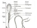
Amytosis. Amytosis occurs by dividing the nucleus, and subsequently the cytoplasm. During amytosis, the nucleolus lengthens, re-lacing, and then the nucleus also stretches. In some cases, a septum appears in the nucleus, which divides it into two parts. Nuclear division is sometimes accompanied by division of the cytoplasm (Fig. 1.75).

Rice. 1.75. Amytosis. Reproduction of amoeba:
a - 0 min; b - 6 minutes; c - 8 minutes; d - 13 minutes; d - 18 minutes; - 21 minutes
There are several forms of amitose: uniform, when two equal nuclei are formed; uneven, when uneven nuclei are formed; fragmentation, when a nucleus breaks up into many small nuclei of the same or different size.
Thus, amytosis is a division that occurs without spiralization without the formation of chromosomes and a division spindle. Or there is a preliminary synthesis of DNA before the onset of amitose and how it is distributed between daughter nuclei is unknown. Sometimes, when certain cells are divided, mitosis alternates with amytosis.
Amytosis is a kind of separation that is sometimes observed during normal cell activity, but mainly in case of dysfunction, often under the influence of radiation or exposure to other harmful factors. It is inherent in highly differentiated cells. Amytosis is less common than mitosis and plays a secondary role in cell division in the vast majority of living organisms.
Mitosis- a method of indirect division of somatic cells.
During mitosis, the cell goes through a series of successive phases, as a result of which each daughter cell receives the same set of chromosomes as in the mother cell.
Mitosis is divided into four main phases: prophase, metaphase, anaphase, and telophase. Prophase- the longest stage of mitosis, during which chromatin condensation occurs, as a result of which X-shaped chromosomes, consisting of two chromatids (daughter chromosomes), become visible. At the same time, the nucleolus disappears, the centrioles diverge to the poles of the cell, and an achromatin spindle (division spindle) of microtubules begins to form. At the end of prophase, the nuclear envelope disintegrates into separate bubbles.
V metaphase chromosomes line up along the cell's equator with their centromeres, to which microtubules of a fully formed division spindle are attached. At this stage of division, the chromosomes are the most dense and have a characteristic shape, which makes it possible to study the karyotype.
V anaphase there is a rapid DNA replication in centromeres, as a result of which chromosomes are cleaved and chromatids diverge to the poles of the cell, stretched by microtubules. The distribution of chromatids must be absolutely equal, since it is this process that ensures the maintenance of the constancy of the number of chromosomes in the cells of the body.
On the stage telophase daughter chromosomes collect at the poles, despiralize, around them nuclear membranes are formed from bubbles, and nucleoli appear in the newly formed nuclei.
After the division of the nucleus, division of the cytoplasm occurs - cytokinesis, during which there is a more or less uniform distribution of all organelles of the mother cell.
Thus, as a result of mitosis, two daughter cells are formed from one mother cell, each of which is a genetic copy of the mother cell (2n2c).
In sick, damaged, aging cells and specialized tissues of the body, a slightly different process of division can occur - amitosis. Amitosis is called the direct division of eukaryotic cells, in which the formation of genetically equivalent cells does not occur, since the cellular components are unevenly distributed. It is found in plants in the endosperm, and in animals in the liver, cartilage and cornea of the eye.
Meiosis. Phases of meiosis
Meiosis is a method of indirect division of primary germ cells (2n2c), as a result of which haploid cells (1n1c), most often sex cells, are formed.
Unlike mitosis, meiosis consists of two successive cell divisions, each of which is preceded by interphase. The first division of meiosis (meiosis I) is called reduction, since in this case the number of chromosomes is halved, and the second division (meiosis II) - equational, since in its process the number of chromosomes is preserved.
Interphase I proceeds like the interphase of mitosis. Meiosis I is divided into four phases: prophase I, metaphase I, anaphase I, and telophase I. B prophase I two important processes take place - conjugation and crossing over. Conjugation is the process of fusion of homologous (paired) chromosomes along their entire length. The pairs of chromosomes formed during conjugation are retained until the end of metaphase I.
Crossover- mutual exchange of homologous regions of homologous chromosomes. As a result of crossing over, the chromosomes received by the body from both parents acquire new combinations of genes, which leads to the appearance of genetically diverse offspring. At the end of prophase I, as in the prophase of mitosis, the nucleolus disappears, the centrioles diverge to the poles of the cell, and the nuclear envelope disintegrates.
V metaphase I pairs of chromosomes are aligned along the equator of the cell; spindle microtubules are attached to their centromeres.
V anaphase I whole homologous chromosomes, consisting of two chromatids, diverge to the poles.
V telophase I around the clusters of chromosomes at the poles of the cell, nuclear membranes are formed, nucleoli are formed.
Cytokinesis I provides separation of the cytoplasm of daughter cells.
The daughter cells (1n2c) formed as a result of meiosis I are genetically heterogeneous, since their chromosomes, which randomly diverge to the poles of the cell, contain dissimilar genes.
Interphase II very short, since there is no DNA doubling in it, that is, there is no S-period.
Meiosis II also divided into four phases: prophase II, metaphase II, anaphase II, and telophase II. V prophase II the same processes proceed as in prophase I, with the exception of conjugation and crossing over.
V metaphase II chromosomes are located along the equator of the cell.
V anaphase II chromosomes are split in centromeres and chromatids are already stretched to the poles.
V telophase II nuclear membranes and nucleoli are formed around the clusters of daughter chromosomes.
After cytokinesis II the genetic formula of all four daughter cells is 1n1c, but they all have a different set of genes, which is the result of crossing over and a random combination of the chromosomes of the maternal and paternal organisms in the daughter cells.
Types of somatic cell division
Mitosis - indirect cell division, which results in the formation of two cells with evenly distributed genetic material.
Amitosis - direct cell division in half, which does not provide an even distribution of genetic material between daughter cells.
Endomitosis - the process of DNA duplication, accompanied by a multiple doubling of chromosomes without dividing the cytoplasm.
Polities - an increase in the amount of DNA, without an increase in the number of chromosomes. Chromosomes become gigantic.
Cell cycle- this is the existence of a cell from the moment of its formation by division of the mother cell to its own division or death.
Mitotic cycle- a complex of events occurring in the process of preparing a cell for division and during the division itself.
The biological significance of mitosis is that as a result, two daughter cells are formed with a set of chromosomes identical to the set of the parent cell. Phases of mitosis - interphase (G - presynthetic, S - synthetic, G - postsynthetic periods), prophase, metaphase, anaphase, telophase.
Chromosomes in a dividing cell are shaped like straight or curved rods. Each chromosome is divided into two arms by a primary constriction, or centromere. Depending on the location of the primary constriction, three types of chromosomes are distinguished: equal arms, or metacentric, unequal arms, or submetacentric and acrocentric (with one long and second very short arm). Some chromosomes have a secondary constriction (nucleolar organizer). In this part of the chromosome, a nucleolus is formed in the interphase nucleus. The metaphase chromosome consists of two chromatids - spirally twisted filaments interconnected in the sphere of the primary constriction. When cell division is completed, chromatids of each chromosome enter different cells and are transformed into independent chromosomes. The main chemical components of chromosomes are DNA (about 40%) and proteins (about 60%). The chromosomes also include RNA, lipids, carbohydrates, metal ions.
The number of chromosomes in the cells of each type of organism is constant. The set of chromosomes in germ cells is called haploid and is denoted by the Latin letter n. The set of paired chromosomes in somatic cells is called diploid and is designated 2n. The set of chromosomes in the cells of organisms belonging to the same species is characterized by a certain size, shape, number and is called a karyotype. All chromosomes in a cell can be divided into two groups - autosomes, or non-sex chromosomes, and sex chromosomes - heterochromosomes. Heterochromosomes determine the sex characteristics of the organism. The human karyotype is represented by 46 chromosomes, of which 44 are autosomes and two sex chromosomes.
Types of division of somatic cells - concept and types. Classification and features of the category "Types of division of somatic cells" 2015, 2017-2018.
The most universal way of somatic cell division, i.e. cells of the body (from the Greek. soma - body), is mitosis. This type of cell division was first described by the German histologist W. Fleming in 1882, who observed the emergence and described the behavior of filamentous structures in the nucleus during division.
From here comes the name of the division process - mitosis (from the Greek mitos - thread).
During mitotic division, the cell nucleus undergoes a series of strictly ordered sequential changes with the formation of specific filamentous structures. In mitosis, several phases are distinguished: prophase, prometaphase, metaphase, anaphase and telophase (Fig. II.2).
Prophase is the first stage of preparation for division. In prophase, the reticular structure of the nucleus gradually turns into visible (chromosomal) filaments due to spiralization, shortening and thickening of chromosomes. During this period, the double nature of chromosomes can be observed, because each chromosome appears to be doubled longitudinally. These halves of chromosomes (the result of reduplication (duplication) of chromosomes in phase 3), called sister chromatin, are held together by one common region - the centromere. The divergence of the centrioles to the poles and the formation of a fission spindle (2n4с) begins.
In the prometaphase, the spiralization of chromosomal filaments continues, the nuclear envelope disappears, the karyolymph and cytoplasm are mixed with the formation of mixoplasm, which facilitates the movement of chromosomes to the equatorial plane of the cell (2n4c).
In metaphase, all chromosomes are located in the equator of the cell, forming the so-called "metaphase plate". At the metaphase stage, chromosomes have the shortest length, since at this time they are most strongly spiralized and condensed. This stage is most suitable for counting the number of chromosomes in a cell, studying and describing their structure, determining sizes, etc. The arrangement of chromosomes in relation to each other is random. The division spindle is fully formed and the spindle filaments attach to the centromeres of the chromosomes (2n4c).
| Anaphase refers to the next phase of mitosis, when chromosome centromeres divide. The spindle filaments pull apart sister chromatids, which from this moment can be called daughter chromosomes, to different poles of the cell. This ensures a consistent and accurate distribution of chromosomal material into daughter cells (2n2c). In the telophase, the daughter chromosomes are despiralized and gradually lose their visible individuality. A nuclear envelope is formed, a symmetrical division of the cell body begins with the formation of two independent cells (2n2c), each of which enters the O, interphase period. And the cycle repeats again. The biological significance of mitosis is as follows. 1. Events occurring during mitosis lead to the formation of two he - | Rice. II.2. Scheme of mitotic cell division: a - interphase; 6, c, d, e - different stages of prophase; f, g - prometaphase; h, i - metaphase; k - anaphase; l, m ~ telophase; and - the formation of two daughter cells |
not physically identical daughter cells, each of which contains exact copies of the genetic material of the ancestor (mother) cell.
2. Mitosis ensures the growth and development of the organism in the embryonic and postembryonic periods. An adult's body consists of approximately 1014 cells, which requires approximately 47 cell division cycles of a single sperm-fertilized egg (zygote).
3. Mitosis is a universal, evolutionarily fixed mechanism of regeneration, that is, the restoration of lost or functionally obsolete cells of the body.
More on topic II.Z. MITOSIS - SOMATIC CELL DIVISION:
- 3. Immortality Becomes Reality (1999) interview with Doctor of Technical Sciences, Senior Scientist of NASA, Professor Alexander Bolonkin
- METHODS FOR GENETIC RESEARCH OF DEVELOPMENTAL DISORDERS. PRENATAL DIAGNOSTICS. ACCOUNTING OF DATA ON GENETIC AND PRENOTAL FACTORS IN PSYCHOLOGICAL ANALYSIS AND DIAGNOSIS.
Cell cycle- the period of a cell's life from the moment of its formation by division of the maternal cell to its own division.
Methods for dividing somatic cells:
1) division in two, or binary;
2) amitosis - direct division;
3) mitosis - indirect division;
4) meiosis - reduction division.
Division in two, or binary characteristic of prokaryotic cells (bacteria), in which there is nucleoid- the genetic apparatus of the bacterial cell (bacterial chromosome). It is a circular DNA molecule that is not linked to histones. The nucleoid is usually located in the center of the cell and is not delimited by its membrane from the contents of the cell. Nucleoid division occurs after the completion of DNA replication. The divergence of daughter DNA is provided by the growth of the cell membrane. Before cell division, DNA is doubled and 2 circular DNA molecules are formed. Then the cell membrane grows into the cytoplasm, is embedded between 2 DNA molecules and divides the cell in two.
Amitosis - direct division the interphase nucleus of the cell by constriction, in which the formation of a spindle of division does not occur. In amitosis, the nucleus divides, and the cytoplasm may remain undivided. In this case, the chromosomes are unevenly distributed. Through amitosis, cells are divided in which pathological processes occur, for example, cells of malignant tumors. In humans and animals, cells of the liver, cartilage tissue, and the cornea of the eye divide amitotically. In plants, endosperm cells divide amitotically. Signs characterizing amitosis:
1) division of the nucleus can occur without division of the cytoplasm;
2) it is found in specialized cells (in the cells of cartilage tissue, the cornea of the eye);
3) the cell in which amitosis has occurred is not capable of mitosis.
Mitosis is the main type of eukaryotic cell division.
Mitosis is indirect division of somatic cells of eukaryotic organisms, in which the daughter nuclei carry the same number of chromosomes as the parent cell. Mitosis provides an increase in the number of cells in the body, growth, and regeneration processes. Chistyakov described some of the phases of mitosis in the spores of the sycamore and horsetail. Then mitosis was studied in detail by a German botanist, E. Strasburger (1876–1879) - in plant cells and a German cytologist, V. Flemming (1882) - in animal cells.
Mitotic cycle- a set of processes occurring in the cell during its preparation for division and during its division.
The mitotic cycle is subdivided into interphase and mitosis.(fig. 26). Interphase- the time interval between cell divisions. The interphase, in turn, is subdivided into three phases - G 1, S, G 2.
In the postmitotic (presynthetic) period - phase G 1 the cell is being prepared for DNA duplication: intensive cell growth; active biosynthesis of RNA, proteins, lipids, carbohydrates, ATP and enzymes.
In the synthetic period - phase S, which lasts 6–8 hours, the main process is carried out - DNA replication (chromosome doubling). DNA synthesis method - replication, or self-doubling molecules DNA. In the course of replication, hereditary information is transmitted from the mother's DNA to the daughter's DNA by means of its exact reproduction. As a result of DNA replication, each chromosome is duplicated and consists of two chromatids. Chromatids are connected in the centromeric region.
In the premitotic (postsynthetic) period - phase G 2 lasting from 2 to 6 hours, there is: duplication of organelles; synthesis of proteins, lipids, carbohydrates, ATP synthesis; proteins are synthesized, which are necessary for the formation of microtubules of the fission spindle.
Rice... 26. Diagram of the mitotic cycle
An organelle, the cell center (centrosome), takes part in the division of animal cells. It is a non-membrane organelle located near the nucleus in the cytoplasm of the cell. The cell center is involved in the formation of the division spindle during cell reproduction. Chromosomes in interphase are doubled, and, entering mitosis, they consist of two sister chroimatids. Mitosis (M) is divided into 4 phases: prophase, metaphase, anaphase and telophase(fig. 27).
Prophase - the stage of mitosis, during which the condensation of chromosomes occurs, the disintegration of the nucleoli, the fission spindle begins to form . V prophase each chromosome consists of two chromatids connected to each other at the centromere. At the end of prophase, the nucleolus disappears, the centrioles diverge to the poles of the cell. A mitotic spindle, consisting of microtubules, arises.
Metaphase- the stage of mitosis, in which chromosomes line up at the equator of the spindle, forming a metaphase plate. At the beginning metaphases the nuclear envelope collapses. Each chromosome is attached by its central region (centromere) to one of the microtubules. There is also a kinetochore, which is located near the centromere and regulates the location and direction of movement of chromosomes. In metaphase, chromosomes are located in the equatorial region of the cell, forming a metaphase plate.
Chromatids are clearly distinguishable during the metaphase of mitosis, when the chromosome consists of two chromatids.
Anaphase - the stage of mitosis, characterized by the divergence of sister chromatids to the opposite poles of the cell. This is the shortest stage of mitosis. After division of the centromere, chromatids diverge into daughter nuclei and become independent chromosomes.
The movement of chromosomes is carried out thanks to the kinetochore and spindle filaments, which contract and stretch chromatids from the equator to the poles of the cell
Telophase- the stage of mitosis, characterized by the formation of daughter nuclei. At the poles, the chromosome cells despiralize and take the form of long filaments, which is characteristic of a nondividing nucleus. Daughter nuclei are formed, and in them - nucleoli. In the daughter nuclei, a nuclear envelope, nucleoplasm, is formed. Throughout the telophase, cytokinesis- division of the cytoplasm, as a result of which two identical daughter cells are separated from each other. They are a genetic copy of the mother's cell and contain a diploid set of chromosomes - 2nc.

Rice. 27. Mitotic phases of an animal cell : A – B prophase; G - prometaphase; D - metaphase; E - anaphase; G - telophase; 3– cytokinesis
The biological significance of mitosis... Mitosis ensures the genetic continuity of cell generations, genetic stability, that is, the species constancy of the number of chromosomes in cells.
Mitotic index(m) is the ratio of the number of cells undergoing mitosis in the tissue to the total number of cells in the tissue or culture. The mitotic index is determined by the formula m = N m / N, where N m is the number of cells undergoing mitosis in the tissue, and N is the total number of tissue cells (1000 cells). Each tissue has its own mitotic index. Its higher indicators are typical for the growth layer of the skin (0.7), the apical and lateral meristems (0.7), the epithelium of the small intestine (0.78), red bone marrow cells (0.74), and lower - for the skeletal muscle tissue (0.0001) and nervous tissue (0.0001).
Meiosis
Meiosis is the process of division of diploid cells of the gonads, during which reduction division is observed, leading to a halving of the number of chromosomes in daughter cells and equalizing division, leading to the formation of gametes. Meiosis was discovered by W. Flemming in 1882 in animals, and E. Strasburger in 1888 revealed a reduction in the number of chromosomes in plants.
Interphase of meiosis. In the interphase, there is a doubling of DNA molecules in the synthetic period. This doubles the chromosomes. Each chromosome contains 2 chromatids (2n2c).
1. The first division of meiosis
Prophase 1... Chromosomes doubled in interphase enter prophase 1.
Therefore, at the beginning of prophase, the chromosomes are doubled (diploid set) and each of them contains 2 chromatids (2n2c). Then the processes (fig. 28) of conjugation and crossing over are carried out. In prophase-1, stages are distinguished: leptotene, zygotene, pachytene, diplotene, diakinesis.
Chromosome conjugation is a process of pairwise temporary convergence of homologous chromosomes. Leptotene- the stage of fine filaments. On the zygotene stages homologous chromosomes come together in pairs and form tetrads - structures of four chromatids, or bivalents. Due to conjugation, each bivalent consists of 4 sister chromatids. The formula for the genetic material is 2n4c.
Crossingover is the crossing of homologous chromosomes or chromatids, accompanied by the exchange of the corresponding sections between chromatids (the process of recombination). On the pachytene stages in bivalents, crossing over occurs: mutual exchange of identical sections along the length of homologous chromosomes, chiasmata are formed - places of crossing of chromosomes. Since each chiasmus corresponds to one crossing-over event, in which two non-sister chromatids are involved, the intensity of the crossing-over process can be judged by the number of chiasmas. In the human chromosome set, the number of chiasmas ranges from 35 to 66. The exchange of regions between non-sister chromatids of neighboring chromosomes - (non-sister exchange) or between sister chromatids - within one chromosome (sister exchange) is possible.
The genetic consequence of crossing over is gene recombination, genetically heterogeneous material is formed, genetic differences arise between chromatids, which provides wide genetic variability of gametes. On the diplotene stages the notebook complex collapses. Homologues repel each other. Diakinesis- the stage completing the prophase of meiosis-1, transitional to metaphase-1. Bivalents are shortened, the nucleus is destroyed, and a fission spindle begins to form.
Metaphase 1... Bivalents, already genetically heterogeneous, are located in 2 layers along the equator of the cell.
Anaphase 1... In anaphase, chromosomes, consisting of 2 chromatids, diverge to the poles, i.e., halves of bivalents diverge. This process is called reduction division, as a result of which two cells are formed, which contain one chromosome, but each chromosome consists of two chromatids. A haploid set of chromosomes is being formed. Therefore, the formula of the genetic material in anaphase-1 has the form - n2c).
Telophase 1... 2 cells are formed with a haploid set of chromosomes and a doubled amount of DNA. The fission spindle is destroyed. The nuclear envelope appears. At the end of telophase 1, cytokinesis occurs (division of the cytoplasm using a constriction); in addition, dyads are formed, i.e. each cell contains 2 sister chromatids connected by a centromere.
So, after the first meiotic division, the cell contains a haploid set of chromosomes, and each chromosome consists of two chromatids.
2. The second division of meiosis - equalizing division (mitosis of meiosis)... Between the first and second divisions of meiosis, there is a period - interkinesis... Unlike interphase, DNA is not replicated in interkinesis, and chromosome duplication does not occur.
The second division of meiosis includes the same phases as the first division - phase-2, metaphase-2, anaphase-2, telophase-2.
In prophase-2 and metaphase-2 of meiosis, two chromatids are still preserved in each chromosome. In prophase II of meiosis, the chromosome set of a cell can be written in the form of the formula 1 n 2 c (n is the number of chromosomes, c is the number of chromatids).
In anaphase-2, sister chromatids diverge to the poles of the cell, and each of them becomes an independent chromosome. As a result of the divergence of chromatids to the poles of the cell, equalizing division.
In telophase -2, the genetic material formula is n c.

Rice. 28 ... Stages of meiosis. Chromosome behavior. The paternal chromosomes are colored black, the maternal chromosomes white.
Thus, meiosis consists of two successive divisions (reduction and equalization). Before the first division of meiosis, in the interphase, DNA synthesis occurs, as a result of which there will be two chromatids in each chromosome (single DNA replication - 2n2c). Reduction division ends with the formation of two cells containing a haploid set of chromosomes, consisting of two chromatids (1n2c). Before the second division, there is no interphase in meiosis. Therefore, the second division is not preceded by DNA synthesis and chromosome doubling. As a result of equalizing division (mitosis of meiosis), 4 haploid genetically dissimilar cells are formed from one initial diploid cell of the gonad. After equalizing division the formula of the genetic material has the form - 1n1c.
The biological significance of meiosis consists: 1) in the formation of genetically diverse material, due to crossing over; 2) in the diversity of species, since meiosis serves as the basis for the combinative variability of organisms; 3) in the formation of gametes participating in sexual reproduction; 4) in maintaining the genetic constancy of species.