The largest bacterium in the world. Bacteria The largest bacteria in size do not exceed
Dwarfs and giants among bacteria
Bacteria are the smallest living organisms, which are the most common form of life on Earth. Conventional bacteria about 10 times the smallest of the human cell. Their size is about 0.5 microns, and you can only see them with a microscope. However, in the world of bacteria, it turns out, also has its own dwarfs and giants. One of these giants is the bacterium Epulopiscium Fishelsoni, the dimensions of which reach half a millimeter! That is, it reaches the size of the sizes of grains or grains of salt and can be seen with her unarmed look.
With the help of sulfur pearls, nature invented an amazing solution to the problem of critical size: hollow bacteria. Inside there is a huge container, 50 times more than cytoplasm, live part of the cell. Like orange peel, cellulose surrounds a living part of the cavity.
Bacteria settled in the world with various fantastic ways. Of all creatures, often forgotten unicellites are the most successful - and still often used by people to overestimate themselves as a crown of evolution. Bacteria live in the kidney stones of people and in the intestines of worms, in the air, in boiling geysers and in the Ice of Antarctica. Some bring suffering, such as plague, cholera or tuberculosis around the world, others help plants grow or people digest, others feed on oil, seas are contaminated, some are even resistant to strong radioactivity.
Epulopiscium reproduction
In the Cornwell Academy, studies were conducted aimed at determining the causes of such large sizes. As it turned out, bacteria stores 85,000 copies of DNA. For comparison, only 3 copies are contained in human cells. This cute creation lives in the digestive tract of tropical reef fish Acanthurus Nigrofuscus (Surgeon Fish).
Surnery pearl plays an important role in the natural cycle of Matter Namibia, and this role formally forced her giantism. It feeds on sulfur compounds, abundant in sediment, which is their home. In order to digest sulfur, bacteria, like metabolism of animals, depend on oxygen - nitrates are urgently needed. But this does not exist in a hostile sauce in which Tiomargarita Namibiensis lives.
This dilemma did not break the simplest, but made it a giant: every few months when the storm beats the water, rich in nitrates, also penetrates the bacteria into depths. Sulfuric pearl can now store the precious nitrate in his cavity, which he is in abundance for a short time; It manages stocks, like a dive, which takes off the compressed air in depths.
The usual views of bacteria are very small and primitive, they have no organs and food occurs through the shell. Nutrients are evenly distributed over the body of bacteria, so they should be small. Unlike them, EPULOPISCIUM repeatedly copies its DNA, evenly, along the shell distributes copies, and they are powered sufficiently. Such a structure gives her the possibility of an instant response to external stimuli. Unlike the remaining bacteria and the method of its division. If ordinary bacteria is simply divided in half, then it grows in itself two cells, which after her death just come out.
Since the largest bacterium on Earth can also store sulfur, it can last months without food - the first Namibian pearl, and then simply stops the air and is waiting for the best times. Today we know that Namibian Pearl of Sulfur not only has many close relatives in other marine areas, but also plays an important role in ecology: these bacteria can cause rock formation with high phosphorus content. This reduces the amount of phosphate in sea water, so that it is no longer available as a nutrient for other living beings.
Namibian sulfuric pearl
However, even this, not a small bacterium, can not be compared with the largest bacterium in the worldwhich is considered Thiomargarita NamibiesisFor another Namibian Sulfur Pearl, a gram-negative nautical bacterium, opened in 1997. It not only consists of only one cell, but at the same time, she has no supporting skeleton as well as Eukarotov. Thiomargarita dimensions reach 0.75-1 mm, which allows you to see it with a naked eye.
Thus, the formation of these breeds opposes the excessive enrichment of oceans with phosphate. Most bacteria are usually very small and can only be detected by a microscope. But gigantic forms appeared in several bacteria groups. They are more than hundreds of times more than ordinary bacteria, and are easily recognized by the naked eye. The largest known bacteria belong to the group of serobacteria. These bacteria can be recognized by bright gray inclusions of sulfur, which cause oxidation of sulfur bacteria with sulfide to sulfur and further sulfate for energy production.

According to the type of metabolism of thyomargaritis, it is an organism that receives energy as a result of restorative and oxidative reactions and can use nitrate as the final object receiving electrons. The cells of the Namibian sulfuric pearl are stationary, and therefore the content of nitrate can fluctuate. Thiomargarita can store nitrate in a vacuole that occupies about 98% of the entire cell. With a low nitrate concentration, its contents are used for breathing. Sulphides are oxidized to nitrates to sulfur, which is assembled in the inner medium of bacteria in the form of small granules than and the pearl color of tyomargartes is explained.
For this, they use either oxygen or nitrate. Breathing with nitrate is also the cause of an unusual size. Cells of giant bacteria consist mainly of large enclosed in the vacuole membranes in which they can store nitrate with high concentration.
Keeping nitrate for breathing and sulfur as an energy source, gigantic bacteria can long survive in adverse external conditions. 
Before Namibia, the seabed contains much more sulphides than in other coastal areas, which, obviously benefits this giant with its corresponding large reservoir of nitrates. In addition, especially soft seabed Namibia regularly twists large-scale methane flashes. Since its opening 14 years ago, these bacteria gained fame and were included in the Guinness Book of Records, as well as depicted on Namibian Mark.
Research Tiomargiti
Studies conducted not so long ago showed that Thiomargarita Namibiesis may not be bonded, but an optional body receiving energy without the presence of oxygen. It is capable of oxygen breathing, if this gas is enough. Another distinctive feature of this bacterium is the possibility of a Palinitomic division occurring without an increase in intermediate growth. This process is used by Thiomargarita Namibiesis in stressful states caused by starvation.
Of course, after opening in Namibia, Toyargarita began in other sulphide-rich marine areas, and indeed, very similar bacteria could be found elsewhere, but nowhere in this number and with so many different forms as near Namibia. Only recently it was possible to genetically explore this variety of manifestations. In addition, two other previously unknown genus were discovered, which are now called Tiopilla and Tyofiz.
Sulfur bacteria and phosphorus cycle
Although he was also discovered on the seabed off the coast of Chile and Costa Rica, it is found there only as a single camera and does not create typical pearl necklaces, which Tyomargarita is obliged to be his name.

In huge cells, serobacteria for storing substances is enough space. Not only sulfur for power supply and nitrate as an oxidizing agent, but phosphate can accumulate the cell as a kind of energy storage as a polyphosphate in large quantities. In coastal areas where a particularly large amount of sulfur bacteria lives, rocks with high phosphorus content, so-called phosphorites are also formed.

Bacterry was opened in the bottom sediments of the equalized outskirts of the mainland, near the Namibian coast, Heid Schulz, a German biologist and its colleagues in 1997, and in 2005, in the cold winches of the Gulf of Mexican Bay, discovered a close strain that is a confirmation of the widespread distribution of Namibian sulfuric pearls. .
In the ancient rocks, which come from marine, coastal areas, you can often find fossils, the form of which resembles sulfur bacteria. All together, this suggests that for a long time, large sulfur bacteria could play a direct role in the phosphoric cycle of the sea, which favors the formation of phosphorites. Now the question arises on the formation of phosphorites, since this process reduces the amount of dissolved phosphate available in seawater as a nutrient for all living organisms.
Victor Ostrovsky, samogo.net
Bacteria - the first "residents" of our planet. These primitive nuclear-free microorganisms, most of which consist only of one cell, subsequently gave rise to other, more complex forms of life. Scientists investigated more than ten thousand of their species, but about a million more unexplored remain uneasy. The standard size of the micromir representative: 0.5-5 microns, but the largest bacterium has a size of more than 700 microns.
Therefore, an increase in phosphorus formation means less growth for all organisms in the long run. In fact, it seems that there is a direct link between the formation of phosphite and large sulfur bacteria. As a result, the mineral apatite is rich in phosphorus, and the first step towards the formation of phosphorites is made.

The seabed at the shores of Namibia is so rich in phosphorites that they are even useful as raw materials for the fertilizer industry. We suspect that similar mechanisms also apply to tyomargite.
Bacteria - an oldest living form on Earth
Bacteria may have spherical, spiral, spherical shapes. They can be found everywhere, they are thick inhabit water, soil, acidic environments, radioactive sources. Scientists find living unicellular microorganisms in the conditions of permafrost and in the erupting lava of volcanoes. You can see them thanks to the microscope, but some bacteria grow to gigantic sizes, completely changing the representation of a person about the micrometer.
It is still unknown why sulphide causes phosphate emissions. In fact, however, it can be noted that both today and in the history of the Earth, phosphorites were formed in a strong sulfide seabed. Therefore, we suspect that these and similar bacteria play an important role in the phosphorus cycle in the sea and probably contributed to the formation of phosphorite in the geological past. What advice gives an expert on health if we ask her questions about how easy and inexpensive to avoid breeding bacteria? "Hand washing", Dr. Eckerli, British hygiene specialist.
In the end, pathogens especially love to appear and often appear where they do not expect them. It is not surprising that 65% of all colds, 50% of all diarrheal diseases and 80% of all gastrointestinal diseases associated with food products fall into "clean" households. Not in the bathroom, but in the kitchen. In most households, the probability of detection of fecal bacteria is 200 times higher.
- Thiomargarita Namibiesis, Namibian sulfuric pearl - the so-called bacteria from famous man. To see it, do not need a microscope, it is 750 microns. The microme giant was discovered by German scientists in the bottom waters during the expedition on the Russian scientific vessel.

- Epulopiscium Fishelsoni lives in the intestine of the fish-surgeon and has a length of 700 microns. The volume of this bacterium is 2000 times higher than the volume of the microorganism of standard sizes. Initially large unicellic was found inside fish surgeons inhabiting the Red Sea, but after it was found in other types of fish in the area of \u200b\u200bthe Great Barrier Reef.
- Spioctuettes - bacteria with long, spiral cells. Very moving. Live in water, in the soil or in another nutritious environment for them. Many spirochetes are the pathogens of serious human diseases, other varieties are saprophitis - decompose an extreme organic. These bacteria can grow to a length of 250 μm.
- Cyanobacteria are ancient microorganisms. Scientists have found their livelihoods whose age is more than 3.5 billion years. These unicellites are part of the ocean plankton and produce 20-40% oxygen on Earth. Spirulina is dried, grind and added to food. Oxygen photosynthesis is characteristic of algae and higher plants. Cyanobacteria is the only single-cellular, which in the process of photosynthesis isolated oxygen. It is thanks to cyanobacteria in the Earth's atmosphere, a large stock of oxygen appeared. The width of the cells in these bacteria varies from 0.5 to 100 microns.

- Actinomycetes inhabit the intestines of most invertebrates. Their diameter is 0.4-1.5 microns. There are pathogenic forms of actinomycetes living in dental and in the respiratory tract of man. Thanks to actinomycetes, man also feels a specific "smell of rain".
- BEGGIATOA ALBA. The proteobacteria of this kind inhabit places rich in gray, fresh rivers and the sea. The size of these bacteria is 10x50 microns.
- Azotobacter has a diameter of 1-2 microns, lives in weakly alkaline or neutral media, plays a large role in the nitrogen cycle, increases the soil fertility and stimulates the growth of plants.
- Mycoplasma Mycoides is the causative agent of pulmonary diseases from cows and goats. These cells have a size of 0.25-0.75 microns. Bacteria do not have a rigid shell, from the external environment they are protected only by the cytoplasmic membrane. The genome of this type of bacteria is one of the easiest.

Archaei are not bacteria, but as they, as they, consist of a single cell. These unicellites were allocated near the thermal underwater sources, inside the oil wells and under the icy surface of the Northern Alaska regions. Archaei have their own evolution of development and differ from other forms of life with some biochemical features. The average size of the archaeus is 1 μm.
Create an immune system - and clean it regularly
Good immune protection is basically intestinal. So good intestinal protection is responsible for our health. Therefore, it is advisable to build an intestinal flora with a good diet. Liquid and hygienic conditions should be obtained for the remaining 20 percent. The most dirtiest household items: kitchen sponges and rags, cutting boards, kitchen countertops, drains, door handles and toothbrushes.
Wet and warm - the perfect climate for breeding. In addition, bacteria are very easily transported from one place to another with textiles. It is best to use individual textile products and often replace them. It is regularly dried: most bacterial strains cannot survive in dry conditions. Good advice: You can disinfect the sponge, wash them in the dishwasher.
Theoretically, the most minimal size of a unicellular microorganism: 0.15-0.20 μm. With a smaller size of the cell will not be able to reproduce itself like, as biopolymers in the desired composition and in the required quantity are not placed in it.
The role of bacteria in nature
In the human body coexist more than a million species of different single-cell microorganisms. Some of them are extremely helpful, others can apply an irreparable damage to health. The first "portion" of bacteria baby receives at birth - during passing through the generic pathways of the mother and in the first minutes after delivery.
Cuts and cracks on the boards provide a large nutrient medium for bacteria. Again, be careful that there are no cross-contaminants: do not use raw meat and raw fish without disinfection. So that the cutting board was completely clean, it is recommended to use this cleaning agent: Mix 1 teaspoon of a chlorine bleach with 200 ml of water. Drain the board, let it dry. You can also deliver cutting boards into a dishwasher.
The biggest problem: Clean the work surfaces only seemingly clean textiles. If you use the same dirty fabrics and kitchen sponges for various dishes, it increases the risk of microbes. Regular disinfection helps. Even the drains provide bacteria with a humid climate. You get them clean with soda or food soda and toothbrush. Thus, stains, stubborn dirt and even smells can easily be sent to flight. Plums can also be regularly cured.

If the child appears on the light of the cesarean section, the kid's body is populated with microorganisms for him. As a result, it decreases natural immunity, the risk of allergic reactions increases. By three years most of the microbiome of the child is formed. Each person has its own unique set of inhabiting its microorganisms.
From hand to hand: bacteria love door handles. If the member is still sick, mini pests are even happier. Especially in this case: regularly wash your hands. Antibacterial soap should be avoided anyway, because these are real shells that kill all bacterial strains. Natural soap is a healthier alternative.
Various bacterial strains
You must change every three months. Not only because of bacteria, also because you break the brushes over time. Despite all the "home confusion" described: bacteria are not bad by themselves. There are good and bad bacterial strains, and most people can easily cope with both strains. Normal households set up a healthy bacterial flora.
Bacteria are used by man in the production of drugs and food products. They split organic compounds, cleaning them and turning dirty drains into harmless water. Soil microorganisms produce nitrogen compounds necessary for plant growth. Unicellites are actively recycled by the organic and carry out a circulation of substances in nature, which is the basis of life on our planet.
Bacteria is the most ancient group of organisms from now existing on Earth. The first bacteria appeared, probably more than 3.5 billion years ago and for almost a billion years have been the only living creatures on our planet. Since these were the first representatives of wildlife, their body had a primitive structure.
Over time, their structure was complicated, but also the bacteria is considered the most primitive single-cell organisms. Interestingly, some bacteria and now have now retain the primitive features of their ancient ancestors. This is observed in bacteria living in hot sulfur sources and oxless flashes at the bottom of the reservoirs.
Most bacteria colorless. Only a few are painted in purple or in a green color. But the colonies of many bacteria have a bright color, which is caused by the separation of the painted substance into the environment or pigmenting of cells.
The plaque of the world of bacteria was Anthony Levenguk - the Dutch natural backs of the 17th century, which first created the perfect magnifying glass microscope, increasing items 160-270 times.
Bacteria refer to prokarytams and isolated into a separate kingdom - bacteria.
Body shape
Bacteria are numerous and varied organisms. They differ in shape.
| Bacteria name | Shape bateria | Image of bacteria |
| Cockki. | Sharo-shaped | |
| Bacillus |  | Chopkoid |
| Vibrio | Semiconde | |
| Spirillum |  | Spiraloid |
| Streptococci |  | Cockkk chain |
| Staphilococci |  | Breakdi Cockkn. |
| Diplococci | Two round bacteria enclosed in one mucous capsule |
Methods of movement
Among bacteria there are movable and fixed forms. Movable moves due to wave-like cuts or with the help of flagellas (twisted screw threads), which consist of a special flask of flagellin. The flagellas may be one or more. They are located in some bacteria at one end of the cell, others - on two or all over the surface.

But the movement is inherent in many other bacteria that there are no flavors. Thus, bacteria covered with mucus outside are capable of sliding movement.
Some devoid harvesters of aquatic and soil bacteria in the cytoplasm there are gas vacuoles. The cell can be 40-60 vacuoles. Each of them is filled with gas (presumably - nitrogen). Adjusting the amount of gas in vacuoles, water bacteria can be immersed in the thickness of the water or rise to its surface, and soil bacteria - move in soil capillaries.
Habitat
Due to the simplicity of the organization and unpretentiousness of the bacteria are widespread in nature. The bacteria were found everywhere: in a drop of even the most pure spring water, in the tillage grains, in the air, on the rocks, in the polar snow, the sands of the desert, at the ocean day, in the huge depth of oil and even in the water of hot springs with a temperature of about 80ºС. They live on plants, fruits, in various animals and in humans in the intestine, oral cavity, on the limbs, on the body surface.
Bacteria are the smallest and most numerous living beings. Thanks to small sizes, they easily penetrate any cracks, slits, pores. Very hardy and adapted to various conditions of existence. Turn the drying, strong cold, heating up to 90 ° C, without losing the viability.
There is practically no place on earth where bacteria would not meet, but in different quantities. The living conditions of bacteria are diverse. One of these requires air oxygen, others do not need it and are able to live in an oxygenous medium.
In the air: bacteria rise to the upper atmosphere of up to 30 km. and more.
Especially many of them in the soil. In 1, the soil may contain hundreds of millions of bacteria.
In water: in the surface layers of water of open reservoirs. Useful water bacteria mineralize organic residues.
In living organisms: pathogenic bacteria fall into the body from the external environment, but only in favorable conditions causing the disease. Symbiotic live in digestion organs, helping to split and absorb food, vitamins synthesize.
External structure
The bacteria cell is dressed by a special dense sheath - a cell wall that performs a protective and reference function, and also gives bacteria a constant characteristic of her form. The cell wall of the bacteria resembles a vegetable cell shell. It permeates: through her nutrients freely pass into the cage, and the metabolic products are entering the environment. Often over the cell wall in the bacteria, an additional protective layer of mucus - capsule is produced. The thickness of the capsule can increase the diameter of the cell itself many times, but maybe very small. Capsule is not a mandatory part of the cell, it is formed depending on the conditions in which bacteria fall. It protects the bacterium from drying.
On the surface of some bacteria there are long flagellas (one, two or many) or short thin veins. The length of flags can many times to exceed the bacterium bodies. With the help of flagella and vigor, bacteria move.

Internal structure
Inside the bacteria cell is a thick fixed cytoplasm. It has a layered structure, there are no vacuoles, therefore various proteins (enzymes) and spare nutrients are placed in the substance of the cytoplasm. Bacteria cells do not have a kernel. In the central part of their cells, a substance serving hereditary information is concentrated. Bacteria, - nucleic acid - DNA. But this substance is not decorated in the core.
The internal organization of the bacterial cell is complex and has its own specific features. The cytoplasm is separated from the cell wall of the cytoplasmic membrane. In the cytoplasm there is a basic substance, or matrix, ribosomes and a small number of membrane structures that perform a variety of functions (mitochondrial analogues, an endoplasmic network, a Golgi apparatus). In the cytoplasm of bacteria cells often contain granules of various shapes and sizes. Granules can consist of compounds that serve as a source of energy and carbon. In the bacterial cell there are and fat droplets.
In the central part of the cell, the nuclear substance is localized - DNA, not degraded from the cytoplasm of the membrane. This is an analogue of the nucleus - nucleoid. The nucleoid does not have a membrane, a nuclear fuel and a set of chromosomes.
Methods of nutrition
Bacteria observed different ways of nutrition. Among them are autotrophic and heterotrophs. Avtotrophs are organisms capable of independently form organic substances for their power.
Plants need nitrogen, but they themselves absorb air nitrogen. Some bacteria connect the nitrogen molecule contained in the air with other molecules, resulting in substances available for plants.
These bacteria settle in the cells of young roots, which leads to formation on the roots of thickening, called non-naval. Such tubers are formed on the roots of plants of the family of legumes and some other plants.
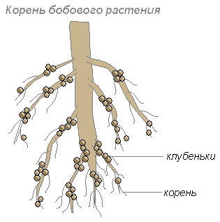
The roots give carbohydrate bacteria, and the bacteria roots are such nitrogen substances that can be assimilated by the plant. Their cohabitation is mutually beneficial.
The roots of plants are distinguished by many organic substances (sugar, amino acids and others), which are powered by bacteria. Therefore, in a layer of soil surrounding the roots, especially many bacteria are settled. These bacteria turn outmended plant residues into a substance available for the plant. This layer of the soil is called the rhizosphere.
There are several hypotheses about penetration of nodule bacteria in the root fabric:
- through damage to the epidermal and cow fabric;
- through root hairs;
- only through a young cell shell;
- thanks to bacteria satellites producing pectinolytic enzymes;
- due to the stimulation of the synthesis of in-indolyluxusic acid from tryptophan, always existing in the root discharge of plants.
The process of introducing nodule bacteria into the root fabric consists of two phases:
- infection of root hairs;
- the process of the formation of the tuber.
In most cases, the introduced cell, actively multiplies, forms the so-called infectious threads and is already in the form of such threads moved to the tissue of the plant. Nodule bacteria that came out of the infectious thread continue to multiply into the host fabric.

Fulled vegetable cells, filling with rapidly multiply cells, begin to share hard. The connection of the young tuber with the root of the leggings is carried out thanks to vascular fibrous beams. During the functioning of the tubers are usually dense. By the time of the manifestation of the optimal activity, the muscles acquire a pink color (thanks to the pigment LeggoLobin). Only those bacteria that contain legglobin are capable of fixing nitrogen.
The bacteria of the tubers create dozens and hundreds of kilograms of nitrogen fertilizers on the hectare of soil.
Metabolism
Bacteria differ from each other metabolism. In some, it goes with the participation of oxygen, others - without his participation.
Most bacteria feed on ready-made organic substances. Only some of them (blue-green, or cyanobacteria) are capable of creating organic substances from inorganic. They played an important role in the accumulation of oxygen in the atmosphere of the Earth.
Bacteria absorb substances from outside, tear their molecules to pieces, from these parts they collect their shell and replenish their contents (so they grow), and unnecessary molecules are ejected out. The shell and membrane of the bacteria allows it to absorb only the necessary substances.
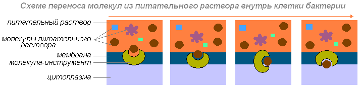
If the shell and membrane bacteria were completely impenetrable, no substances would fall into the cage. If they were permeable for all substances, the contents of the cell would be mixed with the medium with a solution in which bacterium lives. For the survival of the bacteria, a shell is necessary, which the necessary substances skips, and unnecessary - no.
Bacteria absorbs nourishing substances nearby. What happens later? If she can move independently (moving a flagellum or pushing back the mucus), then it moves until the necessary substances find.

If it cannot move, it is waiting for the diffusion (the ability of the molecules of a substance to penetrate into the thick of the molecules of another substance) will not bring the necessary molecules to it.
Bacteria in aggregate with other groups of microorganisms perform tremendous chemical work. Turning various compounds, they get energy and nutrients needed for their lively. The metabolic processes, methods of extracting energy and the need for materials for the construction of substances of their body in bacteria are diverse.
Other bacteria All carbon needs needed to synthesize organic matter bodies are satisfying due to inorganic compounds. They are called autotrophs. Auto-flow bacteria are able to synthesize organic substances from inorganic. Among them are distinguished:
Chemosynthesis
The use of radiant energy is the most important, but not the only way to create an organic substance from carbon dioxide and water. Bacteria are known, which are not used as a source of energy for such synthesis, and the energy of chemical bonds occurring in the cells of organisms during the oxidation of some inorganic compounds - sulfide, sulfur, ammonia, hydrogen, nitric acid, acidic compounds of iron and manganese. The organic substance formed using this chemical energy is used to build cells of their body. Therefore, such a process is called chemosynthesis.
The most important group of chemosynthetic microorganisms is nitrifying bacteria. These bacteria live in the soil and carry out the oxidation of ammonia formed during the rotation of organic residues to nitric acid. The latter, reacts with the mineral compounds of the soil, is converted into nitric acid salts. This process takes place in two phases.
Jamming is converting zakuzny iron into the oxide. The formed iron hydroxide settles and forms the so-called marsh iron ore.
Some microorganisms exist due to the oxidation of molecular hydrogen, thereby ensuring the authotrophic power method.
A characteristic feature of hydrogen bacteria is the ability to switch to a heterotrophic lifestyle while providing them with organic compounds and the absence of hydrogen.
Thus, chemoavtotrophs are typical autotrophic, as the necessary organic compounds are independently synthesized from inorganic substances, and do not take them in the finished form from other organisms as heterotrophs. From phototrophic plants, chemoavtotrophic bacteria differ in complete independence from light as an energy source.
Bacterial photosynthesis
Some pigment-containing serobacteria (purple, green), containing specific pigments - bacteriochlorophylls, are able to absorb solar energy, with which hydrogen sulfide in their organisms split and gives hydrogen atoms to restore the corresponding compounds. This process has a lot in common with photosynthesis and is only distinguished by the fact that purple and green bacteria donor hydrogen is hydrogen sulfide (occasionally - carboxylic acids), and in green plants - water. For those and other cleavage and transfer of hydrogen due to the energy of the absorbed sunlight.
Such bacterial photosynthesis, which occurs without the release of oxygen, is called photoreduction. The photo generation of carbon dioxide is associated with the transfer of hydrogen not from water, but from hydrogen sulfide:
6SO 2 + 12N 2 S + HV → C6H 12 O 6 + 12S \u003d 6N 2 O
The biological importance of chemosynthesis and bacterial photosynthesis across the planet is relatively small. Only chemosynthetic bacteria play a significant role in the process of sulfur circulation in nature. Absorbing green plants in the form of salts of sulfuric acid, sulfur is restored and included in the composition of protein molecules. Next, in the destruction of dead vegetative and animal residues, sulfur with sulfide, which is oxidized by sulfide sulfide, which is oxidized by sulfur-free sulfur (or sulfuric acid), sulfite-free in soil. Chemo and photoauthotrophic bacteria are essential in the cycle of nitrogen and sulfur.
Sporing
Inside the bacterial cell disputes are formed. In the process of sporing, the bacterial cell undergoes a number of biochemical processes. It decreases the amount of free water, enzymatic activity is reduced. This ensures the stability of the dispute to the adverse conditions of the outer environment (high temperature, high saline concentration, drying, etc.). Sponges are typical only by a small group of bacteria.

Disputes - not the mandatory stage of the life cycle of bacteria. Sponge formation begins only with a lack of nutrients or accumulation of exchange products. Bacteria in the form of an argument can be at rest for a long time. Spores of bacteria withstand long-term boiling and very long-term industrialization. Upon the occurrence of favorable conditions, the dispute germinates and becomes viable. Conference bacteria is a device for survival in adverse conditions.
Reproduction
Bacteria is multiplied by the division of one cell into two. Having achieved a certain size, bacterium is divided into two identical bacteria. Then each of them begins to eat, grows, it is divided and so on.

After lengthening the cell, the transverse partition is gradually formed, and then daughter cells diverge; In many bacteria, under certain conditions, the cells after division remains associated with the characteristic groups. In this case, depending on the direction of the plane of division and the number of divisions, different forms occur. The reproduction of the kill is found in bacteria as an exception.
Under favorable conditions, cell dividing in many bacteria occurs every 20-30 minutes. With such a rapid reproduction, the offspring of one bacterium in 5 days is able to form a mass that can fill all seas and oceans. A simple calculation shows that 72 generations (720,000,000,000,000,000,000 cells) can be formed during the day. If we translate into the weight - 4720 tons. However, this does not occur in nature, since most bacteria die quickly under the action of sunlight, during drying, disadvantage of food, heating to 65-100ºС, as a result of the struggle between the species, etc.
Bacteria (1), absorbed enough food, increases in size (2) and begins to prepare for reproduction (cell division). Its DNA (in the bacteria of the DNA molecule is closed in the ring) doubles (bacterium produces a copy of this molecule). Both DNA (3,4) molecules are attached to the bacterium wall and the bacterial lengthening is diverged on the parties (5.6). First divides nucleotide, then cytoplasm.
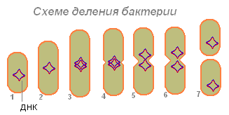
After the discrepancy between the two DNA molecules on the bacteria, a hauling appears, which gradually separates the body of the bacterium into two parts, in each of which there is a DNA molecule (7).
It happens (in a hay stick), two bacteria sticks out, and a jumper is formed between them (1.2).

On the jumper DNA from one bacterium is transferred to another (3). Called in one bacterium, DNA molecules are flying, sticking together in some places (4), after which they exchange areas (5).
The role of bacteria in nature
Crooked
Bacteria is the most important link of the total cycle of substances in nature. Plants create complex organic substances from carbon dioxide, water and mineral soil salts. These substances are returned to the soil with extreme mushrooms, plants and animal corpses. Bacteria decompose complex substances on simple, which again use plants.
Bacteria destroy complex organic substances of dead plants and animal corpses, allocating living organisms and different garbage. Feeding by these organic substances, the saprophistic bacteria of rotting turn them into humus. These are peculiar socitars of our planet. Thus, bacteria are actively involved in the cycle of substances in nature.
Soil formation
Since bacteria are spread almost everywhere and are found in a huge amount, they largely determine the various processes occurring in nature. In the fall, the leaves of trees and shrubs fall, the overhead feces of herbs die, fall out of the old branches, falling from time to time the trunks of old trees fall. All this gradually turns into humus. In 1 cm 3. The surface layer of forest soil is contained hundreds of millions of saprophistic soil bacteria of several species. These bacteria are converted by humus in various minerals that can be absorbed from the soil roots of plants.
Some soil bacteria can absorb nitrogen from the air using it in the processes of vital activity. These nitrogen-free bacteria live independently or settled in the roots of legume plants. Penetrating in the roots of legumes, these bacteria cause the growth of the roots cells and the formation of nodule on them.
These bacteria isolated nitrogen compounds that use plants. Bacteria from plants are obtained carbohydrates and mineral salts. Thus, there is a close relationship between the beam and nodule bacteria, useful for both one and other organism. This phenomenon is called symbiosis.
Thanks to symbiosis with nodule bacteria, legumes enrich the soil with nitrogen, contributing to raising the harvest.
Distribution in nature
Microorganisms are distributed everywhere. The exception is only the crater of the active volcanoes and small sites in the epicenter of the blown atomic bombs. No low temperatures of Antarctic, nor boiling jets of geysers, nor saturated solutions of salts in hydrochloric basins, nor strong insolation of mountain peaks, nor the hard irradiation of atomic reactors interfere with the existence and development of microflora. All living beings constantly interact with microorganisms, being often not only by their storage facilities, but also by distributors. Microorganisms are the aborigines of our planet, actively mastering the most incredible natural substrates.
Microflora soil
The number of bacteria in the soil is extremely large - hundreds of millions and billion individuals in 1 gram. In the soil they are much larger than in water and air. The total number of bacteria in soil changes. The number of bacteria depends on the type of soil, their states, the depth of the layer.
On the surface of soil particles, microorganisms are located small microcoltones (20-100 cells each). Often they develop in the thickness of the bunches of organic matter, on the living and dying roots of plants, in thin capillaries and inside the lumps.
The soil microflora is very diverse. There are different physiological groups of bacteria: bacteria of rotting, nitrifying, nitrofixing, serobacteria, etc. Among them are aerobes and anaerobes, disputes and not disputes. Microflora is one of the factors of soil formation.
The area of \u200b\u200bdevelopment of microorganisms in the soil is a zone adjacent to the roots of living plants. It is called the rhizosphere, and the aggregate of microorganisms contained in it - the rhizosphere microflora.
Microflora reservoirs
Water is a natural environment where microorganisms develop in large numbers. The bulk of them enters the water from the soil. A factor determining the amount of bacteria in water, the presence of nutrients in it. The most pure are the water of artesian wells and spring. Very rich in bacteria open reservoirs, rivers. The largest number of bacteria is located in the surface layers of water, closer to the shore. When removing from the shore and increasing depth, the number of bacteria decreases.
Clean water contains 100-200 bacteria in 1 ml., And contaminated - 100-300 thousand and more. Many bacteria in the bottom Ile, especially in the surface layer, where the bacteria form the film. In this film, there are many seroids and ferrucks, which oxidize hydrogen sulfide to sulfuric acid and thereby prevent the fish to the fisher. In Ile more sporing forms, while in the water predominantly dominated.
According to the specified composition of the water microflora is similar to the microflora soil, but there are also specific forms. Destroying various garbage in the water, microorganisms gradually carry out the so-called biological purification of water.
Microflora air
Air microflora is less numerous than soil microflora and water. Bacteria rise into the air with dust, some time can be there, and then settle on the surface of the Earth and die from lack of nutrition or under the action of ultraviolet rays. The number of microorganisms in the air depends on the geographical area, the terrain, the time of year, pollution of dust and others. Each dusting is a carrier of microorganisms. Most of the bacteria in the air over industrial enterprises. Air countryside cleaner. The most clean air over the forests, mountains, snow spaces. The upper air layers contain fewer microbes. In the air microflora, many pigmented and sporing bacteria, which are more resistant than others, to ultraviolet rays.
Microflora of the human body
The human body, even completely healthy, is always a carrier of microflora. When contacting the body of a person with air and soil on clothes and skin, a variety of microorganisms, including pathogenic (tetanus sticks, gas gangrenes, etc.) are seen. Open parts of the human body are most often contaminated. In the hands of intestinal sticks, staphylococcis are found. In the oral cavity there are over 100 types of microbes. The mouth with its temperature, humidity, nutritional residues is an excellent environment for the development of microorganisms.
The stomach has an acidic reaction, so the bulk of microorganisms in it is dying. Starting from the subtle intestine, the reaction becomes alkaline, i.e. Favorable for microbes. In the thick intestines of the microflora is very diverse. Each adult highlights every day with excrement about 18 billion bacteria, i.e. More individuals than people on the globe.
Internal organs that are not connected with the external environment (brain, heart, liver, bladder, etc.), usually free from microbes. In these organs, microbes fall only during the disease.
Bacteria in cycle of substances
Microorganisms in general and bacteria in particular play a large role in biologically important cyphans of substances on Earth, carrying out chemical transformations that are completely inaccessible neither by plants or animals. The various stages of the cycle of elements are carried out by organisms of different types. The existence of each individual group of organisms depends on the chemical transformation of the elements carried out by other groups.
Crack of Nitrogen
The cyclic transformation of nitrogen compounds plays a paramount role in supplying the necessary forms of nitrogen of various food needs of the biosphere organisms. Over 90% of the total nitrogen fixation is due to the metabolic activity of certain bacteria.
Create carbon
The biological conversion of organic carbon into carbon dioxide, accompanied by the reduction of molecular oxygen, requires the joint metabolic activity of a variety of microorganisms. Many aerobic bacteria exercise complete oxidation of organic matter. In aerobic conditions, organic compounds are initially split off by saving, and organic finite fermentation products are further as a result of anaerobic breathing if there are inorganic hydrogen acceptors (nitrate, sulfate or CO 2).
Circular sulfur
For living organisms, sulfur is mainly available in the form of soluble sulfates or reduced organic sulfur compounds.
Crooked Iron.
In some reservoirs with fresh water, reduced iron salts are kept in high concentrations. In such places, a specific bacterial microflora is developing - barrels, oxidizing reduced iron. They participate in the formation of marsh iron ore and water sources rich in iron salts.
Bacteria are the most ancient organisms that appeared about 3.5 billion years ago in Archeye. About 2.5 billion years, they dominated the Earth, forming the biosphere, participated in the formation of an oxygen atmosphere.
Bacteria are one of the most simply arranged living organisms (except for viruses). It is believed that they are the first organisms that appeared on Earth.
In this article, we invite you to a fascinating tour of the list of 25 largest living beings on Earth, ranging from giants according to the microman - viruses, bacteria and ameb to the largest invertebrates, insects, amphibians, birds, reptiles, fish, mammals, plants and Mushrooms.
1. The largest of famous viruses (1.5 μm in length)
You can argue for a long time, whether there are actually viruses with alive organisms - some biologists say yes, others are not so confident. Nevertheless, there is no doubt that Pithovirus. A real giant among the well-known virus science (about 1.5 μm in length), 50 percent more of the nearest record holder - Pandoravirus.. Perhaps you thought that the causative agent of this size as Pithovirus. It is capable of infecting large animals such as elephants, hippos or even people. But do not worry, the virus affects only Ameb, which is not much more than himself.
2. The largest bacterium (more than 0.5 mm in length)

Thiomargarita Namibiesis - Translated Latin means Namibian Sulfur Pearl. This name bacterium received because of the sulfur granules included in the cytoplasm, giving it a brilliant appearance. The size thiomargarita Namibiesis It is more than half a millimeter in width, which makes it possible to consider it with an unarmed eye. Thiomargarita Namibiesis Absolutely harmless to people and animals, as it is a lithotrof (organisms using inorganic substances as oxidized substrates (electron donors)).
3. The largest Ameb on the planet (3 mm in length)

The largest Amebe refers to the family "Chaos". Of course, it is much less monstrous Ameb from comics and science fiction films. But still, this is a real Giant among ameb, which is easy to see the naked eye. Another feature of the world's largest amoeba is the ability to digest small multicellular organisms, bacteria and protists.
4. Heavy beetle (85-110 g)

Despite the fact that Goliath is not the longest beetle in the world, however, taking into account their mass (some individuals weigh more than 100 g), they undoubtedly correspond to their name. The Goliaf Beetle, by weight and size, comparable to the adult mice with sand, in which you have already convinced you see the photo above.
5. The largest spider (body weight up to 175 g)

Terafoz Blonde or Poultryed Goliath is the largest spider of the world, originally from the tropical forests of South America. Considering the legs, the poultry-goliaf body length can reach up to 28 cm, and the weight - until 175. The life span of females of the giants of giants in the wild is up to 25 years, and sexual maturity comes in 3 years. The males were less fortunate, despite the fact that they do not eat the female after the statement of the pairing, like other types of spiders, their life expectancy is shorter - from 3 to 6 years.
6. The largest worm (average length 60-90 cm)

If you are experiencing a strong dislike for worms, then you can alarm the fact of the existence of more than half of the dozen species of giant worms - the biggest of which is an African giant worm, up to 1.5 m long And harmless, as their small fellow. They love to be buried deep in the mud, stay away from people (and other animals), calmly drinking rotten leaves and other decomposing organic substances.
7. The largest amphibian (body weight up to 3 kg)

Goliath is a popular name for the largest representatives of the fauna (see paragraphs No. 4 and No. 5). Goliaf frog lives in the Western Central Part of Africa. The middle weight of the Goliath frog is about 2.5 kg, which is much less than the mass Beelzebufo ampinga. (about 5 kg) - the largest frog living on Earth during the late Cretaceous period.
8. The largest segmental animal (3-4 m, taking into account the legs)

The Japanese crab spider is truly a huge and extremely long animal. The front feet of this representative of arthropods reaches lengths up to 2 m, and torso up to 45 cm. Motley, orange-white color of the exoskeleton serves as an excellent disguise from large marine predators. Like most other strange creatures, the Japanese spider crab is a valuable delicacy in Japan, but recently, it is rarely found in the restaurant menu due to pressure from the defenders of natural resources.
9. The largest flowering plant (diameter up to 1 m)

Fortunately for all of us, the habitat of Rafflesii Arnold is limited to Indonesia, Malaysia, Thailand and the Philippines. You definitely do not meet it in the neighbor garden. :)
10. The largest sponge on the planet (up to 2 m in diameter)

Besides the giant sea sponge (XESTOSPONGIA Muta) The largest of its kind, it is a record holder for life expectancy among invertebrate animals, some individuals live more than 1000 years. Like other types of sponges, xESTOSPONGIA Muta. It is powered by filtering small organisms from sea water.
11. The largest jellyfish (up to 37 m in length)

With a dome diameter of about 2 m and tentacles more than 30 m, the length of the pouring cyanium is comparable with blue whale (see clause number 22). Despite such gigantic sizes, the tentacles of these jellyfish are not a fatal hazard for humans (only painful sensations and rash on the skin). Washed cyania, also performs an important environmental function, providing various types of fish and crustacean shelter under a huge dome.
It is interesting that the fact that the hairy cyania is a favorite source of food for another giant in this list - leathery turtle (see paragraph number 17)
12. The largest flying bird (adult males weigh up to 20 kg)

Given the huge (according to bird standards) body weight - up to 20 kg, the African big darf is against the laws of aerodynamics. This is not the most elegant bird in the world when it comes to flight. In fact, the African big darf, a significant part of life spends on land in the southern part of Africa, loudly buckle and absorbing almost everything that moves. She uses only in cases of extremely danger.
In this regard, the African Big Drop does not differ from even larger Pterosaurov - flying reptiles of the Mesozoic era.
13. The largest representative of the protists (more than 45 m in length)

Many people mistakenly believe that there are only four categories of life - bacteria, plants, mushrooms and animals - but do not forget about primitive eukaryotic organisms, such as chromists. Most likely you will be surprised by the fact that all algae belongs to the protists. The biggest representative of the protists is Macrocystis Pyrifera. - The type of brown algae from the laminarium family, which is capable of growing more than 45 m in length, providing a reliable shelter with many marine organisms.
14. The largest non-flying bird (up to 270 cm in height and weighing up to 156 kg)

If you take globally, the ostrich is not only the largest not flying bird, but in general the biggest bird from now living on Earth. The maximum registered ostrich height is 2.7 m, and the weight is 156 kg. It may seem incredible, but relatively recently (about 200-300 years ago) on the island of Madagascar, the view of the birds - Madagascar epiornis, compared with which the ostrich would seem like chicken. These birds could reach 3-5 m in height and up to 500 kg in weight, which is comparable to the birds of the bird of drromornis (Dromornis) who lived on the planet during the late Miocene period.
15. The biggest snake (mass - 97.5 kg)

Compared to other organisms from this list, the classification of snakes in size is significantly difficult. Even professional naturalists tend to overestimate the sizes of the serpets, which they observed in the wild, since, transportation of large copies for a detailed study is practically no possible. At the same time, most scientists agree that Anaconda is the largest snake of the planet. The largest of the caught Anacond, had a length of 521 cm and a mass of 97.5 kg.
16. The largest representative of bivalve mollusks (more than 200 kg)

Giant Thdakna is the largest view of bivalve mollusks, found in the waters of the Pacific and Indian Oceans. The maximum mass of giant thidaknans is more than 200 kg, and the length of the sink may exceed 1 m. Despite the formidable reputation, the giant mollusk close its sink only in cases of danger, and its size is not enough to swallow the adult.
17. The largest turtle (mass of more than 500 kg)

Leather turtle is a large view of sea turtles living in tropical latitudes. These turtles are equally different from their relatives. The shelf turtle armor consists of small bone plates and is not attached to the skeleton, like other species. In addition to the body structure, the distinctive feature of the leathery turtles is their giant size - the mass of an adult individual may exceed 500 kg.
18. The biggest reptile (weight up to 1000 kg)

According to dinosaur standards, when the largest reptile weighed 100 tons, a rowed crocodile is just a small lizard. Nevertheless, in the world of modern reptiles - these crocodiles are real giants. The length of the body of an adult dog's combed crocodile varies from 3.5 to 6 m, and the mass of 200 to 1000 kg.
19. Largest fish (maximum weight 2235 kg)

A peculiar appearance of an ordinary moon-fish makes it one of the strange inhabitants of the ocean. But these fish are known not only to an eccentric species, as well as their gigantic sizes. The record from the caught copies of an ordinary moon-fish, had a length of 4.26 m and a mass - 2235 kg.
20. The largest land mammals (the average weight of 5 tons)

Mammal from the genus of African elephants, as well as the largest ground animal. The middle mass of the female is 3 tons, and the male - 6 tons. Adult Savan Elephant, is able to eat about 200 kg of vegetation daily and drink up to 200 liters of water.
21. The largest shark (more than 12 m in length)

Oddly enough, but the largest animals of the oceans are usually powered by microscopic organisms. Like a blue whale (see the next item), the whale ration of the shark mainly consists of plankton, small squid and fish. What swings the size of a whale shark, then it is not possible to call exact numbers here. There are various sources that argue about caught giant features more than 20 m in length and weight up to 40 tons. Given the thrust of many fishermen to exaggeration, it is impossible to be 100% confident in this data. Apparently, the more real sizes of the whale shark are 12-14 m in length.
22. The largest marine animal (200 tons)

In fact, blue whale is not only the largest marine animal, but also most likely the largest animal in the history of life on earth, science is not yet known dinosaurs or other reptiles weighing 200 tons. Like a whale shark (see previous item), Blue whale is powered by a microscopic plankton, filtering countless gallons of sea water through the tight plates of the whale TSA. Naturalist scientists believe that adult blue whale consumes 3-4 tons of Krill every day.
23. The largest mushroom (600 tons)

Perhaps in your understanding the biggest mushroom has a leg thick in a pillar and a hat with a roof size of the house, but in reality everyone looks different. Mushroom recordsman, or rather, the colony of mushrooms, which has a common fungne and functions, as a single organism, is located in the forests of the state of Oregon, the United States and refers to the genus of the Opel. The colony covers an area of \u200b\u200b2000 acres and has a total mass of about 600 tons. The age of a giant mushroom, according to the calculations of botany is more than 2400 years.
24. The largest single tree (about 1000 tons)

Giant sequoia - a tree, according to the truth of the giant sizes. The height of the giant sequoia trunk reaches 100 m, with a diameter of 10-12 m, and the calculated weight of the largest trees is about 1000 tons. They also belong to the most ancient organisms on the planet, the rings of one tree in the North-West USA indicated the age of 3500 years.
25. The largest colony of trees (6000 tons)

Like a colony of mushrooms (see clause 23), the largest colony of the poplar aspen "Pando", located in Utah, the United States has a common root system and identical genes. Simply put all the colony trees - clones that occurred from the total ancestor about 80,000 years ago. Unfortunately, at present, Pando in poor condition, slowly fading from drought, diseases and invasions of insects. Botany desperately try to solve the problem, so we hope this colony will be able to flourish at least 80,000 years.
Despite the fact that very obvious eggs of birds and fish Most people eat almost daily, with the words "single-cell organism" seems to be something that can be seen only in the microscope. Indeed, the overwhelming majority of unicellular creatures do not exceed the dimensions in the hundredths of the millimeter, and this is explained by a number of factors. Large living cells are harder to maintain the integrity of the structure, it is more difficult to transport food and waste inside the body, in addition, impressive growth requires a fair energy, which is evolutionary unprofitable.
But the world of microbes is rich in views, old and diverse, therefore is full of exceptions from the rules. And some organisms, which would shift the prefix "Micro", contrary to evolutionary benefits at all. What naturally admire and fascinates.
Infusoria-Trubacch
This freshwater being is like a pipe of an ancient gramophone and grows up to 2 mm in length, so the trumpeter infusoria can be studied without instruments. The simplest genus Sentor is well known to microbes lovers. Two millimeters do not seem supelnin, but many multicellular children of nature occupy a much less place in the habitat and on the slide glasses.
The trumpeter infusoria makes her anatomy in the world in the world. Unlike ordinary eukaryotes, St. Centor contains not one, but several cores. It makes it easier for him daily work to maintain himself in the Spirit. In the case of this infusoria, numerous small cores are responsible for reproduction, and the large kernel - Makronukleus - Heads all the other, playing the role of a sort of brain center.
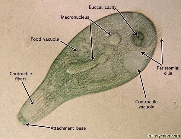
The trumpeter calves is covered with cilia of different lengths. Their friendly movements allow infusories to swim. These colosses of the microcosm feed on, for example, il. The function of the mouth performs the narrow tip of the "pipe". At the same time, some bacteria fall into food, small simplest and even tiny unlucky multicellular.

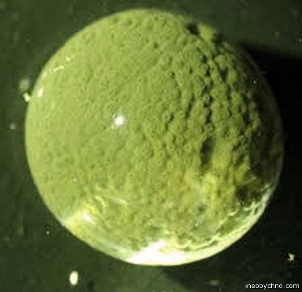
Bahamas thunder
One day, scientists from Texas Uni went to the bottom of the Marine near the Bahamas and found there, in the gloomy depths, dozens of unusual spherical objects with grapes. These objects seemed fixed, but clearly left traces on sand up to half a meter. First, experts thought about some unknown mollusks or even strangely leading cochasts. The truth was amazed, because the mysterious bugs turned out to be a spherical simplest diameter of up to 3 centimeters. Which rolled along the bottom of the sea in the almost zero water temperature.

Bahamas has an amoboid organism having a sink, soft and porous. In the holes in Oyoy, pseudopodia are encouraged, with the help of which thunder moves along the bottom, feeding by the organic matter, which fell along the way.

The discovery of this creature changed some views on the evolution of living beings, since it was previously believed that the first in the Precambrian Starin was learned to crawl into multicellular animals with bilateral symmetry. And traces that leave thresholds are very similar to ancient fossil prints, which are almost 2 billion years old.
Unfortunately, little is known about these balls with cytoplasm, because the living copies of the Live copies are very difficult to deliver into the laboratory. Despite its shells, the simplest fragile and vulnerable. Scientists say that they are much softer grapes, which these giants are microbes are like something.
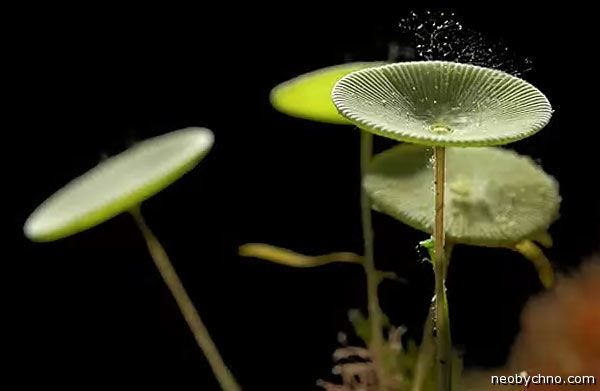
Acetabulary.
Known as "Rusalogy Glass", acetabulary is a unique genus of green algae, similar to the shape of hay mushrooms. These plants of the shallow water of the tropical seas are up to 10 cm long and grow usually by groups, attaching legs to bottom stones and bangs with their light green caps.

Typically, large unicellular creatures have more than one nucleus, which cannot be said about an amazing acetabulary, which spends most of the life with one gigantic container of DNA, located at the heart of its "stem". Only an hour of reproduction is formed additional kernels migrating in the top of the algae, where they turn into spore-like cysts, koi after wintering and complex transformation become young acetabulariors. The life cycle of these colossal warcitis is about three years.

During the experiments conducted for the Nazis money in the 1930s and 40s by the German scientist Joachim Hammerling, it was found that after a transplantation in the same type of algae acetabulary, the source plant begins to form a new hat, transforming into an unusual hybrid.

In addition, the "glass, from which the mermaids drink" perfectly regenerates, being damaged than quite reminds some multicellular views of the world of flora and fauna.
Puzzled Wolonia
Some clich by this funny shallow creature "The Eye of the Sailor", others - simply "algae-bubble". Wolney Puzzy easily grows up to 4 cm in diameter and even more, one organism is one live cage with many nuclei, most often geographically lonely and always similar to polished pebbles of greenish colors. Sometimes on the surface of this unicellular maritime miracle, small "multicellites" are coming.

Despite the biological strangeness and the exotic look of algae, the owners of large marine aquariums do not complain. If the plant is randomly settled, it will capture all the bottom, it is terribly difficult to get rid of it. It is not a deal to put pressure or tear into parts of this living weed. "It is the cellular division of a nuclear filling with its" collection "and breeds.


Kaulerp Tissiste
You might think about it, as if it was some fern, but in essence my plant is much easier. And much more decisively in growth. The fact that the inexperienced diver will seem over the thickets of the underwater flora, in fact, will be one or just a few live cells, "disguised" under complex multicellular bush. These primitive creations are called "Kaulerpa Taxifolia", or just a caulelepa-tree, an amazing creeping stem tissistic. One cell of this green algae with its countless DNA repositors can very quickly distribute almost three meters of styre, which regularly occurs in the Mediterranean Sea, destroying the healthy ecology of the depths. For which Kaulerpa-Christmas tree is recognized as a malicious weed. In California, this "microbe-giant" is generally considered an illegal type.

The Mediterranean variety of tear-tissistic caulelemp, the cells of which reaches a record dimensions, is obliged to person with their pest status. Another half a century ago, this unusual alga in the Mediterranean did not dwell at all. But in the 1970s, a certain aquarium in Germany ordered samples of caulemp from the tropics, but not just for beauty and simple care. The inquisitive Germans subjected to the "Christmas tree" technical bullying. Macrofit irradiated with ultraviolet and treated with chemical mutagens. The result was a single-cell monster, very fast growing and resistant to lower habitat. The cold-resistant and cute algae in 1980 was released in the Mediterranean Sea - someone from the amateur aquarists from Monaco tried.

In four years, it happened inevitable. After flight from the aquarium, Mutoving Kaulerpa victorious occupied the coastal waters of the Mediterranean. Unlike the natural fellow, the mutant cell turned out to be not only aggressive, but also resistant to pollution. In addition, able to regenerate from a piece of size in a centimeter. And poisonous. Attempts to clear from the thickets of Kaulerp, the resort shallow water failed.

Therefore, at the end of the 20th century, the nickname "Alga-killer" was entrenched at the Kaulerpa Taxipolia unicellular organism. The plant is included in the hundreds of the most dangerous invasive species, stop the spread of which - the sacred debt of each non-indifferentian earthland.

Ameba Khaos
Imagine Ameba from the school textbook. Increase it to the size of sesame grains. You will have a Caos Carolinensis creature. Since such simplest changes are constantly changing, then the chaos record holders are capable of stretching to 5 mm in length. Such high-charm unicellites can be fatally hurt, just covering the microscope glass glass.

Despite its impressive sizes, Chaos Carolinensis behaves in the same way as its microscopic relatives, carriers of falselyones. With the help of pseudoenia, the chaos moves, they have enough food. Then the food in vacuoles is digested by alive, and the remnants of the garbage are thrown out of the cell outside. A huge amoeba of microbes of other species is eating, as well as small animals like branchist racks. Chaos will have almost non-stop until it becomes ready for reproduction.

Like the neighbors on the list of the giants of the world of microbes, single-celled chaos has many control centers, simply because one core is not able to control such a massive cell. Depending on the size, Chaos Carolinensis may have up to 1000 cores.

Spirostomum
Speartomum infusoria can be found and unspoken in both fresh and salted waters. And take for some little worm. The extended spirostomet reaches a length of 4 millimeters. Only when looking into the eyepiece of the microscope it becomes clear that this is a moving creature - one large and very long cage covered with a dense fish of the cilia.

Spirostum - the world champion of microbes for the ability to change the volume of the body. Being disturbed, the infusorium can hurt 75% in time less than 1/200 second - faster than any other live cell.

In contrast to the voracious infusorium-trumpets, the spiritomomum does not eat multicellular creatures, but only by bacteria. The giants are multiplied by simply division and do not really like if there are heavy metals in the water, which makes these infusories by the eologists.

Sirirgammina fragile
Another noble candidate for the title of the largest unicellular creature on Earth is a fragile "monster" from class xenophyophophore. In this class of "wearing other people's body" organisms include many inhabitants of the ocean bottom, cytoplasm clutches, building for themselves in the eternal night of fragile wicker "houses" from the remains of other creatures, such as sponges or radilation. Construction glue Cells xenophyophosphor makes themselves, on teams entering chemically from numerous nuclei, which is floating in massive cytoplasm clots. The largest of these bunches reaches 20-centimeter sizes, is eagerly colonized by the worms and wears the species name Syringammina Fragilissima.

Unfortunately, the life and biology of Sirringammines ("sand flute Pan" in translation) is still little studied. Scientists suspect that it feeds on a single-core bacteria, but what the process itself looks like, no one has seen. It is believed that the microbes for their diet of Sirringammin the brigal grown herself inside itself. The reproduction mechanism of these rizarians is also unclear.

Opened fragile deep-sea creatures in 1882, the Scots, the native Northwestern shores. Subsequently, Sirringammin was found on the shelf of the North Africa.

The name of them Legion ...
Among the terrestrial single-celled giants special attention deserved, of course, the metering sludge, the inhabitants of the dead wood. Which initially and for a long time took for mushrooms.

However, the mucus (in particular, the polyphonic fusarium) were not only primitive, but also something much smarter than mushrooms. On the interesting conclusions of Japanese scientists can be found in the material.
Attempts to comply with the genome of a giant sulfur bacterium Achromatium Oxaliferum They gave a paradoxical result: it turned out that each bacterial cell contains not one, but a multitude of differing genomes. The level of intracellular genetic diversity A. Oxaliferum Comparish with a variety of multi-shaped bacterial community. Apparently, the differing chromosomes multiply in different areas of the cytoplasm, subdivided by large calcite inclusions on the set of weakly reporting compartments (compartments). An important role in maintaining internal genetic diversity is played by numerous mobile genetic elements that contribute to gene transfer from chromosome on chromosome. The authors of the discovery suggest that the natural selection of this unique organism is not so much at the level of cells, as at the level of individual compartments within one giant cell.
1. Mysterious bacterium
Giant sulfur bacteria Achromatium Oxaliferumit was discovered in the XIX century, but its biology still remains mysterious - largely because Achromatium is not amenable to cultivation in the laboratory. Achromatium cells can reach 0.125 mm long, which makes it the largest freshwater bacteria (in the seas there are even larger sulfur bacteria, such as Thiomargarita.Is the most older Precambrian embryos turned out to be a bacteria about which? , "Elements", 01/15/2007).
Achromatium Oxaliferum Lives in bottom sediments of fresh lakes, where it is usually found on the border of oxygen and oxygen-free zones, but penetrates in completely oxygen-free layers. Other varieties of (or species) of Achromatium live in mineral springs and in salt precipitation of tidal-tidy marches.
Achromatium gets energy due to the oxidation of hydrogen sulfide, first to sulfur (which is stored in the form of granules in the cytoplasm), and then to sulfates. It is capable of fixing inorganic carbon, but organic compounds can also be absorbed. It is unclear whether he can do only to authotrophic metabolism or he needs an organic feeding.
A unique feature of the achromatium is the presence of numerous large inclusions of colloidal calcite in its cells (Fig. 1). Why it is necessary for bacteria and what role the calcium carbonate is played in its metabolism, it is definitely not known, although there are plausible hypotheses (V. Salman et al., 2015. Calcite-Accumulating Large Sulfur Bacteria of the Genus Achromatium. in Sippewissett Salt Marsh).
The cytoplasm of Achromatium is null in the lumens between calcite granules, which actually divide it into a variety of reported compartments (compartments). Although the compartments and are not fully isolated, the exchange of substance between them seems to be difficult, especially since the prokaryotic is much weaker than in eukaryota, the system of active intracellular transport is developed.
And now it turned out that calcite granules are not the only unique feature of Achromatium. And not even the most striking. In the article published in the journal Nature Communications.German and British biologists reported paradoxical results to which attempts have led to read genomes of individual cells A. Oxaliferum From the bottom sediments of Lake Shtehlin (Stechlin) in the north-east of Germany. These results are so unusual that it is difficult to believe in them, although there are no reason to doubt their accuracy, apparently, no: work is carried out in a methodological relationship very carefully.
2. Confirmation of polyploidity
Although Achromatium, as mentioned, belongs to non-cultivated bacteria, this inconvenience is partly compensated by gigantic cell sizes. They are perfectly visible in the light microscope even with a slight increase, and they can be selected by manually from samples of bottom precipitation (pre-missed through the filter to remove large particles). That is how the authors collected the material for their research. Cells A. Oxaliferumcovered with an organic case, on the surface of which a variety of spripers are sisha - small bacteria. All this concomitant microbiota authors were thoroughly washed off from selected cells to reduce the share of foreign DNA in samples.
To begin with, the researchers painted the achromatium cells with a special fluorescent dye for DNA to understand how much in the cell of the genetic material and how it is distributed. It turned out that DNA molecules are not timed to some one section of the cytoplasm, and form a set (on average about 200 per cell) of local clusters in the lumens between calcite granules (Fig. 1, b, d).
Considering everything that today is known about the major bacteria and their genetic organization, this fact is already enough to consider proven that A. Oxaliferum It is a polyploid, that is, each of its cells contains not one, but many copies of the genome.
However, thoroughly, it is clear that such a huge prokaryotic cell could not do the only copy. It would simply be enough to ensure the entire cage necessary for the synthesis of protein transcripts.
Judging by the fact that the clusters of DNA differ in brightness of fluorescence, these clusters are likely to contain a different amount of chromosomes. Here it is necessary to make a reservation that usually the entire genome of the prokaryotic cell is placed on a single annular chromosome. For Achromatium, this is not proven, but very likely. Therefore, the authors for simplicity use the term "chromosome" as the synonym for the term "one copy of the genome", and we will do the same.
At this stage, nothing sensational was yet discovered. There were those times when everyone thought that the prokaryotic was always or almost always only one ring chromosome in each cell. Today there are already many types of polyploid bacteria and Archey (see, "Elements", 06/14/2016).
3. Methage of the multi-shaped community - in one cell
Miracles began when the authors started the release of DNA from selected and washed cells and to sequencing. Of the 10,000 cells were obtained by a metagen (see Metagenomik), that is, a plurality (about 96 million) short-sized random fragments of chromosomes (rides) belonging to different individuals and in the aggregate giving an idea of \u200b\u200bthe genetic diversity of the population.
The researchers have begun sequencing DNA from individual cells. First, from 27 cells, fragments of the 16S-RDNA gene were isolated, according to which it is customary to classify prokaryotes and according to which the presence of a particular type of microbes usually determine the presence of a sample. Almost all selected fragments belonged to Achromatium (that is, approximately coincided with the sequences of 16S-RRNA achromatium, already available in genetic databases). It follows from this that the DNA studied was not contaminated with the genetic material of some foreign bacteria.
It turned out that every cell A. Oxaliferum,unlike the overwhelming majority of other prokaryotes, contains not one, but several different variants (alleles) of the 16S-RDN gene. It is difficult to determine the exact number of options, because small differences can be explained by sequencing errors, and if you consider "different" only highly different fragments, then the question arises, aside Strongly they should differ. Using the most stringent criteria it turned out that in each cell there are approximately 4-8 different alleles of the 16S-RRNA gene, and this is a minimal assessment, and in fact they are most likely more. This sharply contrasts with a situation characteristic of other polyploid prokaryotes, in which, as a rule, on all chromosomes of one cell, the same variant of this gene is sits.
Moreover, it turned out that alleles of the 16S-RRNA gene, present in the same cell A. Oxaliferumoften form a very far from each other twigs on the general genealogical tree of all the variants of this gene, found (earlier and now) A. Oxaliferum. In other words, 16S-RRNA alleles from one cell are no more relative to each other than alleles taken at random from different cells.
Finally, the authors spent the total sequencing of DNA from six individual cells. For each cell, about 12 million random fragments - rides were read. In the normal situation, this would be enough for an excess, so that with the help of special computer programs to assemble from the rides using their overlapping parts, six very high-quality (that is, read with a very high coating, see Coverage) individual genomes.
But it was not there: although almost all of the Rhyssa was undoubtedly belonged to Achromatium (the admixture of foreign DNA was negligible), read fragments flatly refused to be collected in the genome. Further analysis clarified the cause of failure: it turned out that DNA fragments isolated from each cell actually belong not to one, but a set of quite differently different genomes. In fact, the fact that the authors got from each individual cell is not a genome, but metagen.Such ride sets are usually obtained when analyzing not one organism, but a whole population, which also has a high level of genetic diversity.
This conclusion was confirmed by several independent ways. In particular, dozens of genes are known, which are almost always present in bacterial genomes in a single instance (Single Copy Marker Genes). These single-sourced marker genes are widely used in bioinformatics to verify the quality of assembly of genomes, estimates of the number of species in metagenomic samples and other similar tasks. So, in genomes (or "metagenomas") of individual cells A. Oxaliferum Most of these genes are present in the form of several differing copies. As in the case of 16S-RRNA, the alleles of these single-sources that are in the same cell, as a rule, are not more relative to each other than alleles from different cells. The level of intracellular genetic diversity turned out to be comparable with the level of diversity of the entire population, estimated on the basis of a metagenome of 10,000 cells.
Modern metagenomic already has methods that make it possible to single out fragments from a variety of heterogeneous scraps discussed in the sample, to highlight fragments, with a high probability belonging to the same genome. If there are quite a lot of such fragments, then you can collect a significant part of the genome and even the full genome. It was in this way that the new Nadilip of Archei - Asgardarhei was recently opened and described in detail (see the new Archey Nadilip, to which the ancestors of Eukariot, Elements, 16.01.2017). The authors applied these methods to "metagenomas" of individual cells. A. Oxaliferum. This made it possible to reveal in each "metagen" 3-5 sets of genetic fragments corresponding to, most likely, individual ring genomes (chromosomes). Or rather, each such set corresponds to a whole group of genomes similar to each other. The number of different genomes in each cell A. Oxaliferummost likely more than 3-5.
The level of differences between genomes present in the same cell A. Oxaliferum, approximately corresponds to interspear: bacteria with such a level of differences, as a rule, belong to different types of one kind. In other words, a genetic diversity present in each individual cell A. Oxaliferum,it is comparable not even with a population, but with a multi-shaped community. If the DNA from the single cell of the achromatium was analyzed by modern metagenomic methods "blindly", not knowing that all this DNA comes from one cell, then the analysis would definitely show that there are several types of bacteria in the sample.
4. Intracellular gene transfer
So, U. A. Oxaliferuma fundamentally new thing is found, the unheard of a genetic organization. Of course, the discovery generates a lot of questions, and above all the question "How can it be?!"
We will not consider the most uninteresting option, which is that all this is the result of gross mistakes made by researchers. If so, we will soon find out about this: Nature Communications. "The magazine is serious, the study will want to repeat other teams, so it is unlikely that the refutation will make himself wait long. It is much more interesting to discuss the situation, based on the assumption that the study was carefully conducted and the result is reliable.
In this case, you must first try to find out the reasons for the detected A. Oxaliferumunprecedented intracellular genetic diversity: how it is formed why it is preserved, and how the microbe itself will be wounded to survive. All these questions are very difficult.
In all the other polyploid prokaryotes studied today (including the famous readers of the "elements" of Sulbive Archaei Haloferax Volcanii.) All copies of the genome present in the cell, no matter how much they are, are very similar to each other. Nothing like a tremendous intracellular diversity found A. Oxaliferum,they are not observed. And this is not an accident. Polyploidity gives prokaryotes a number of advantages, however, it contributes to the uncontrolled accumulation of recessive harmful mutations, which in course the account can lead to extinction (for details, see the news polyploidity of eukaryotic ancestors - the key to understanding the origin of mitosis and meiosis, "elements", 14.06.2016).
To avoid the accumulation of mutation cargo, polyploid prokaryotes (and even polyploid plastides of plants) actively use the gene conversion - an asymmetric version of homologous recombination, in which two alleles do not change in places, moving from chromosome on chromosome, as with crosslinic drive, and one of the alleles is replaced by another. This leads to the unification of chromosomes. Thanks to intense gene conversion, harmful mutations are either quickly "rubbed" a unspoken version of the gene, or go to homozygous state, manifest themselves in a phenotype and rejected by selection.
W. A. Oxaliferum The gene conversion and the unification of chromosomes are likely to also occur, but not across the entire cell, and at the level of individual "compartments" - lumens between calcite granules. Therefore, different variants of the genome accumulate in different parts of the cell. The authors checked it with the help of selective staining of different allele variants of the 16S-RRNA gene (see Fluorescent in situ. Hybridization). It turned out that in different parts of the cell, the concentration of different allelic variants really differs.
However, this is still not enough to explain the highest level of intracellular genetic diversity detected from A. Oxaliferum. The authors see its main reason in the high pace of mutagenesis and intracellular genomic rearrangements. Comparison of chromosome fragments from the same cell showed that these chromosomes seem to live a very stormy life: constantly mutate, rebuilt and exchanged areas. W. A. Oxaliferum From Lake Shtehlin, the number of mobile genetic elements has sharply increased compared to other bacteria (including with the nearest relatives - achromatiums from salty marches, in which the level of intracellular diversity, judging by preliminary data, is much lower). The activity of mobile elements contributes to frequent genomic perestroins and the transfer of DNA sections with one chromosome to another. The authors even invented a special term for this: "Intracellular Gene Transfer, IGT", by analogy with all known horizontal gene transfer (HGT).
One of the bright testimonies of frequent rearrangements in chromosomes A. Oxaliferum - The differing order of genes in different versions of the genome, including within the same cell. Even in some conservative (rarely changing during evolution), the operas are sometimes spaced in different sequences on different chromosomes within one cell.
Figure 2 schematically shows the main mechanisms, which, in the opinion of the authors, create and maintain a high level of intracellular genetic diversity A. Oxaliferum.
5. Intracellular selection
Frequent perestroika, intracellular transfer of genes, high pace of mutagenesis - Even if all this and maybe it is bad to explain the high intracellular genetic diversity (and I think that we will not talk about it below), it remains unclear how Achromatium is obscured in such Conditions to maintain viability. After all, the overwhelming majority of non-latheral (affecting the fitness) of mutations and rearrangements should be harmful! Polyploid prokaryotes already have an increased tendency to accumulate mutational goods, and if we also admit ultra-high pace of mutagenesis, it becomes completely incomprehensible, as such a creature, like ahromatium, can exist.
And then the authors push a truly innovative hypothesis. They suggest that the natural selection of the Achromatium acts not so much at the level of whole cells, how much at the level of individual compartments - weakly reporting lumens between calcite granules, in each of which, probably, their genome variants are multiplied.
At first glance, the assumption may seem wild. But if you think about, why not? To do this, it is enough to assume that each chromosome (or every local cluster of similar chromosomes) has a limited "radius of action", that is, proteins encoded in this chromosome are synthesized and operate mainly in its nearest surroundings, and not stirred evenly throughout the cell. Most likely, it is. In this case, those compartments where there are more successful chromosomes (containing a minimum of harmful and maximum mutations), will faster replicating their chromosomes, they will become more, they will begin to spread inside the cell, gradually displacing less successful copies of the genome from neighboring compartments. You can imagine this in principle.
6. Intracellular genetic diversity needs additional explanations
The idea of \u200b\u200bthe intense intracellular selection of genomes, responding to one question (why Achromatium does not die off at such a high pace of mutagenesis), immediately creates another problem. The fact is that thanks to this selection more successful (fasterly replicating) copies of the genome must exhibit inside the cell less successful copies, inevitably reduced In this case, intracellular genetic diversity. The most that we wanted to explain from the very beginning.
Moreover, it is obvious that intracellular genetic diversity should be dramatically decreased at each cell division. Different chromosomes are sitting in different compartments, so in the division, each subsidiary will not receive everything, but only some of the genome variants available from the maternal cell. It can be seen even in Fig. 2.
Intracellular selection Plus Complementation of genomes - two powerful mechanisms that must reduce the inner diversity so quickly that no conceivable (compatible with life) the pace of mutagenesis will not be able to resist this. Thus, intracellular genetic diversity remains unexplained.
Discussing the results obtained, the authors repeatedly refer to our work, which the polyploidity of eukaryotic ancestors is told about the news - the key to understanding the origin of mitosis and meiosis. In particular, they mention that polyploid prokaryotam is very useful to exchange genetic material with other cells. However, they believe that in the life of Achromatium, the intercellular genetic exchange does not play a big role. This is justified by the fact that in the achromatium metagen, although genes were found to absorb DNA from the external environment (transformation, see Transformation), but there are no genes for conjugation (see Bacterial Conjugation).
In my opinion, the genetic architecture of Achromatium indicates not to conjugation, but to more radical methods for mixing genetic material of different individuals, such as the exchange of whole chromosomes and cell fusion. Judging by the data obtained, from a genetic point of view of a cell A. Oxaliferum It is something like a prokaryotic plasody or syncytia, such as those formed as a result of the merger of a set of genetically heterogeneous cells in mucus. Recall that Achromatium - bacteria is non-cultivable, therefore it is possible that some elements of its life cycle (such as periodic fusion of cells) could elude microbiologists.
In favor of the fact that the intracellular genetic diversity of the achromatium is formed not Intracellularly testifies to one of the main facts found by the authors, namely, the alleles of many genes in the same cell form far from each other branches on the phylogenetic tree. If all intracellular diversity of alleles was formed inside the clonically breeding cells that do not change with each other genes, it would be necessary to expect that alleles within the cell will be more relative to each other than alleles from different cells. But the authors convincingly showed that it was not. In general, I would put that the fusion of cells is present in the life cycle of Achromatium. This seems to be the most economical and plausible explanation of the colossal intracellular genetic diversity.
In the final part of the article, the authors hint that the genetic architecture of Achromatium can shed light on the origin of eukaryotes. They formulate it like this: " By the way, Markov and Treaschairs suggested that, like Achromauma from Lake Shtehlin, proto-eukaryot cells could be quickly mutating, diversifying their chromosomes, polyploid bacteria / arches" It is quite right, but we also showed that such a creature could not survive without an intersive interhorganism genetic exchange. Let's hope that further research is spaced light for the remaining riddles of Achromatium.


